
Views: 0 Author: Site Editor Publish Time: 2026-11-05 Origin: Site
Ankle fractures are among the most common fractures of the lower extremity. They frequently result from twisting injuries, falls, sports activities, and high-energy trauma. Among the different patterns of ankle fractures, a trimalleolar ankle fracture is a complex injury involving all three malleoli: the medial malleolus, lateral malleolus, and posterior malleolus.
Because these structures contribute to the stability and alignment of the ankle joint, a trimalleolar fracture can significantly compromise ankle function. The injury may also involve ligament disruption, syndesmotic instability, joint surface damage, and ankle dislocation.
Understanding the fracture pattern, injury mechanism, and degree of articular involvement is essential for selecting an appropriate treatment strategy and restoring ankle stability.
Ankle fractures are commonly classified according to the number of malleoli involved.
The three main categories are:
Unimalleolar fracture: Involves one malleolus, most commonly the lateral malleolus.
Bimalleolar fracture: Involves both the medial and lateral malleoli.
Trimalleolar fracture: Involves the medial, lateral, and posterior malleoli.
Trimalleolar fractures represent a smaller proportion of ankle fractures than unimalleolar and bimalleolar injuries, but they are often associated with greater instability and more complex articular damage.
The ankle joint is formed primarily by the distal tibia, distal fibula, and talus. The three malleoli create the bony structure surrounding the talus.
1. Medial malleolus
The medial malleolus is the distal projection of the tibia on the inner side of the ankle. It contributes to medial ankle stability and provides attachment for the deltoid ligament complex.
2. Lateral malleolus
The lateral malleolus is the distal end of the fibula. It forms the outer wall of the ankle mortise and plays an important role in maintaining the position of the talus. The distal fibula is also closely associated with the syndesmotic ligament complex.
3. Posterior malleolus
The posterior malleolus is the posterior portion of the distal tibia, forming part of the posterior articular surface of the ankle joint. It contributes to the posterior stability of the ankle and provides attachment for the posterior inferior tibiofibular ligament.
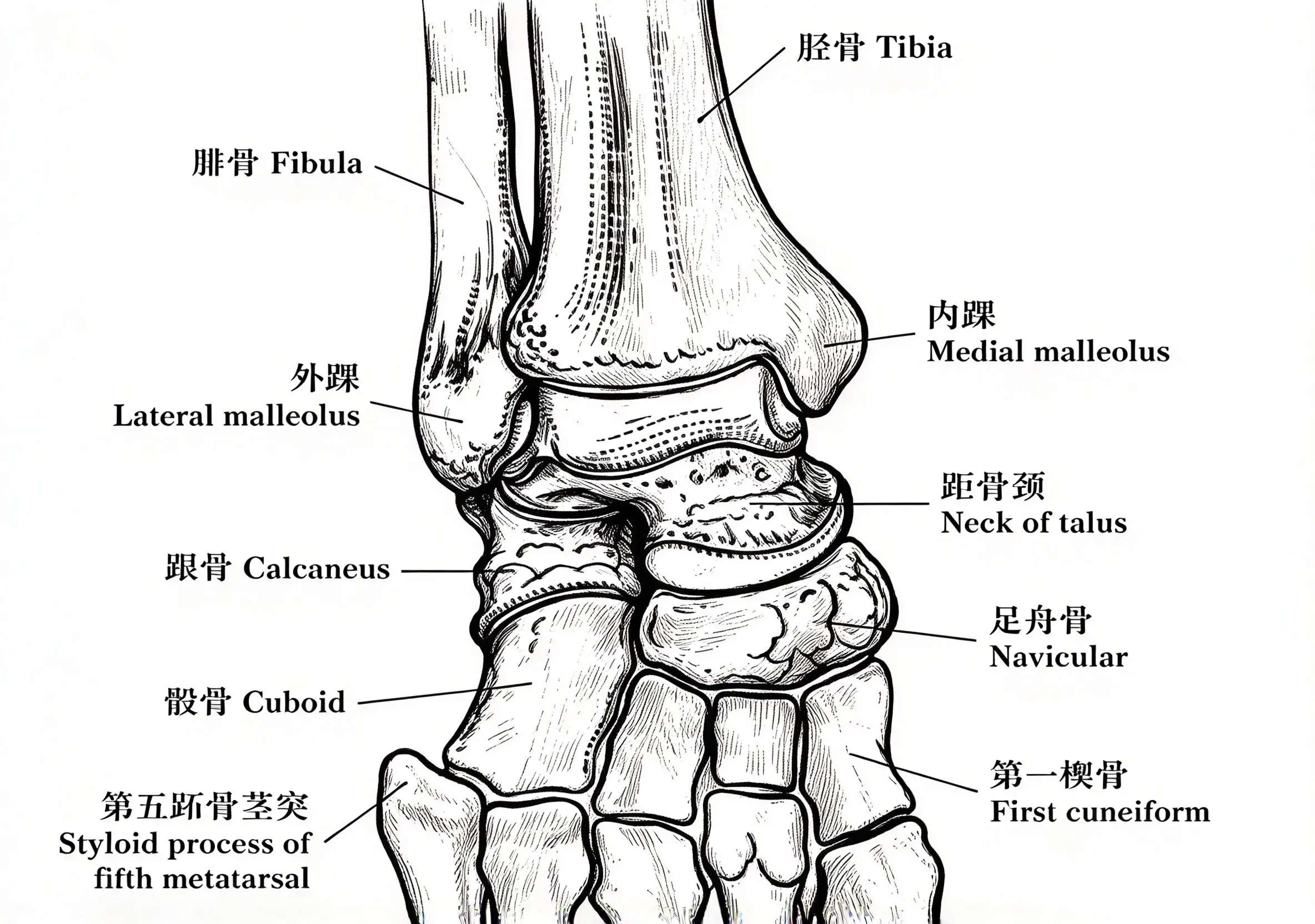
A trimalleolar ankle fracture occurs when all three malleolar structures sustain fractures. Associated ligament injuries may further compromise the ankle mortise, resulting in joint instability, subluxation, or dislocation.
Patients with trimalleolar fractures typically present with significant ankle pain and swelling following an injury.
Common clinical signs and symptoms include:
Swelling: Marked swelling around the ankle, sometimes extending into the foot.
Ecchymosis: Bruising and subcutaneous bleeding around the ankle.
Deformity: The ankle may appear rotated, inverted, or everted, particularly when fracture-dislocation is present.
Tenderness: Localized tenderness over the medial, lateral, and posterior ankle regions.
Restricted movement: Pain and mechanical instability may limit ankle dorsiflexion and plantarflexion.
Difficulty bearing weight: Patients are often unable to stand or walk normally.
In severe cases, an open fracture or ankle fracture-dislocation may occur. These injuries require urgent assessment because of the risk of skin compromise, neurovascular injury, and associated soft-tissue damage.
A complete examination should include:
Inspection of the skin for swelling, bruising, blisters, open wounds, and threatened skin.
Assessment of foot perfusion, including pulses, capillary refill, and skin temperature.
Evaluation of sensory and motor function.
Assessment of tenderness and deformity around the ankle.
Evaluation for associated injuries when the trauma mechanism suggests more extensive damage.
When a fracture-dislocation is present, urgent reduction may be necessary to restore alignment and relieve pressure on the surrounding soft tissues. Neurovascular status should be documented before and after reduction.
Understanding the injury mechanism helps clinicians interpret the fracture pattern and identify associated ligament injuries.
Two important concepts in ankle injury biomechanics are supination and pronation.
Strictly speaking, inversion and eversion are movements primarily associated with the subtalar joint. Supination and pronation describe more complex combined movements involving the foot and ankle.
Supination: A combined movement involving inversion, adduction, and plantarflexion.
Pronation: A combined movement involving eversion, abduction, and dorsiflexion.
During an ankle injury, the foot position and direction of the applied force influence which anatomical structures are damaged.
Supination injuries
Supination injuries can produce tensile forces on the lateral structures and compressive forces on the medial side of the ankle.
Depending on the direction and magnitude of the applied force, the injury may involve the lateral ligaments, distal fibula, medial structures, or posterior malleolus.
Pronation injuries
Pronation injuries can place tensile stress on the medial structures and compressive stress on the lateral side.
These mechanisms may produce medial malleolar fractures, deltoid ligament injuries, fibular fractures, and syndesmotic disruption.
However, the sequence of ligament and bone injury is not determined by tensile or compressive loading alone. The exact fracture pattern depends on the combination of foot position, force direction, rotational movement, and injury energy.
Historically, orthopedic surgeons have used two main characteristics to describe ankle fracture mechanisms:
The position of the foot at the time of injury.
The direction of the external force applied to the ankle.
For example, a supination-external rotation (SER) injury describes an ankle injury in which the foot is supinated and an external rotational force is applied.
These characteristics form the basis of the Lauge-Hansen classification system. They help clinicians understand the potential sequence of structural damage and anticipate associated injuries.
The magnitude of the applied force is also important. Higher-energy trauma can produce more extensive fractures, displacement, soft-tissue damage, and joint instability.
There is no separate universally accepted classification system exclusively for trimalleolar fractures. Instead, these injuries are evaluated using established ankle fracture classification systems.
The three commonly used systems are:
Lauge-Hansen classification
Danis-Weber classification
AO/OTA classification
Each system describes different aspects of the injury and can help guide clinical assessment and treatment planning.
The Lauge-Hansen classification was developed through experimental studies of ankle injury mechanisms.
It is based on two components:
The position of the foot at the time of injury.
The direction of the applied force.
The first term describes foot position, while the second term describes the direction of the force.
The four major injury patterns are:
1. Supination-External Rotation (SER)
This is one of the most common ankle fracture patterns. It typically begins with injury to the anterior inferior tibiofibular ligament, followed by a spiral fracture of the distal fibula at or near the level of the syndesmosis. More severe injuries may involve the posterior malleolus or posterior inferior tibiofibular ligament and the medial structures.
2. Supination-Adduction (SA)
This pattern commonly involves a transverse fracture of the medial malleolus or injury to the deltoid ligament, followed by a vertical fracture of the lateral malleolus when the injury progresses.
3. Pronation-Abduction (PA)
This pattern may involve medial ligament or malleolar injury, syndesmotic disruption, and a fracture of the fibula at or above the syndesmosis.
4. Pronation-External Rotation (PER)
This pattern often involves medial-sided injury, syndesmotic disruption, and a higher fibular fracture. The posterior malleolus may also be involved.
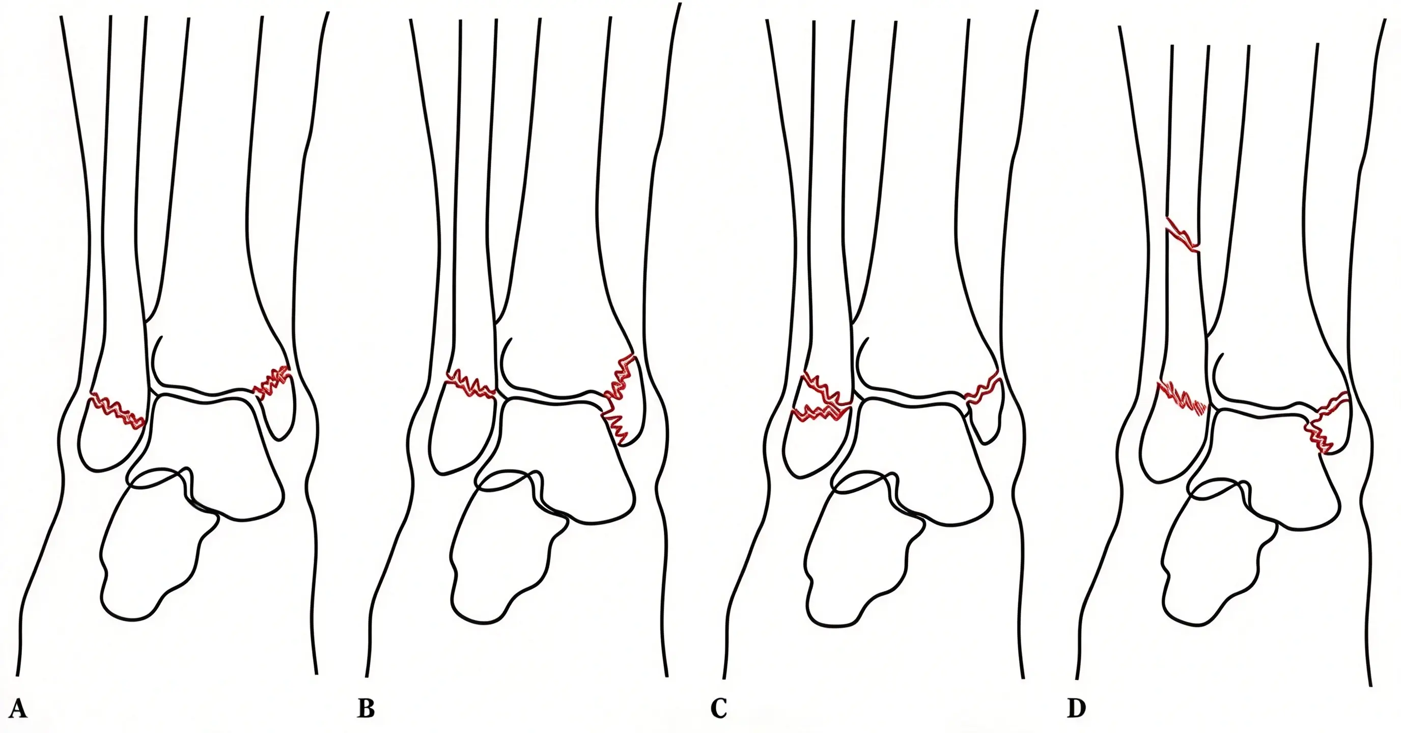
The Lauge-Hansen system is useful for understanding the biomechanics of ankle fractures. However, not every clinical fracture follows the exact sequence described in the original experimental model. Imaging findings and direct clinical assessment remain essential.
The Danis-Weber classification is based primarily on the level of the fibular fracture in relation to the distal tibiofibular syndesmosis.
It divides ankle fractures into three main types.
Weber A: Below the syndesmosis
The fibular fracture occurs below the level of the syndesmosis.
The syndesmosis is generally intact.
The ankle mortise may remain stable if the medial structures are also intact.
Weber B: At the level of the syndesmosis
The fibular fracture occurs at the level of the syndesmosis.
The fracture may be associated with partial or complete syndesmotic injury.
Medial-sided injury may also be present.
Weber C: Above the syndesmosis
The fibular fracture occurs above the syndesmosis.
Syndesmotic disruption is commonly associated.
The injury may involve more extensive damage to the distal tibiofibular structures.
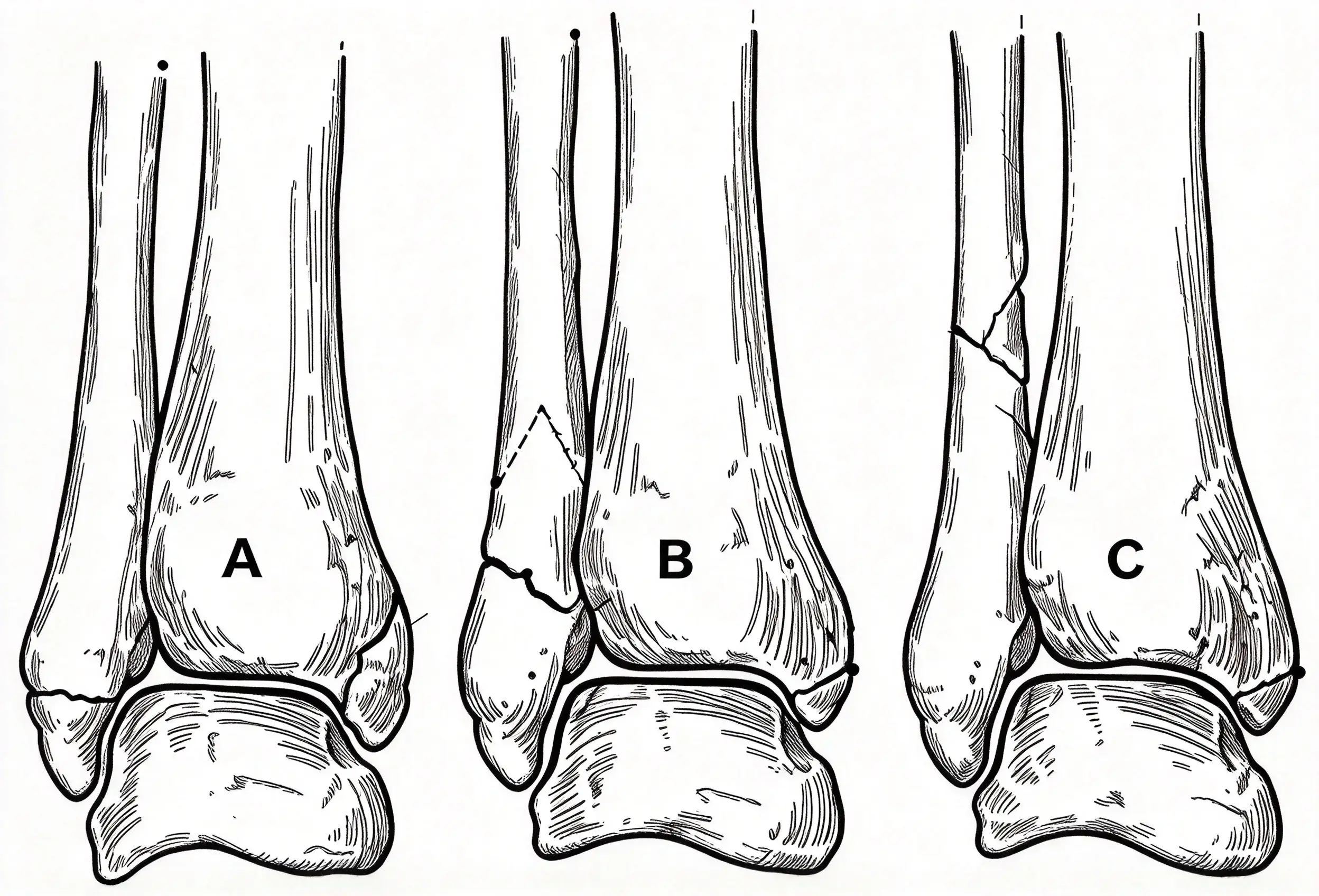
The main advantage of the Danis-Weber system is its simplicity. It provides a straightforward way to describe the fibular fracture level and helps clinicians consider the possibility of syndesmotic instability.
However, the classification does not independently establish whether the ankle is stable. Medial injury, posterior malleolar involvement, and the condition of the syndesmotic ligaments must also be evaluated.
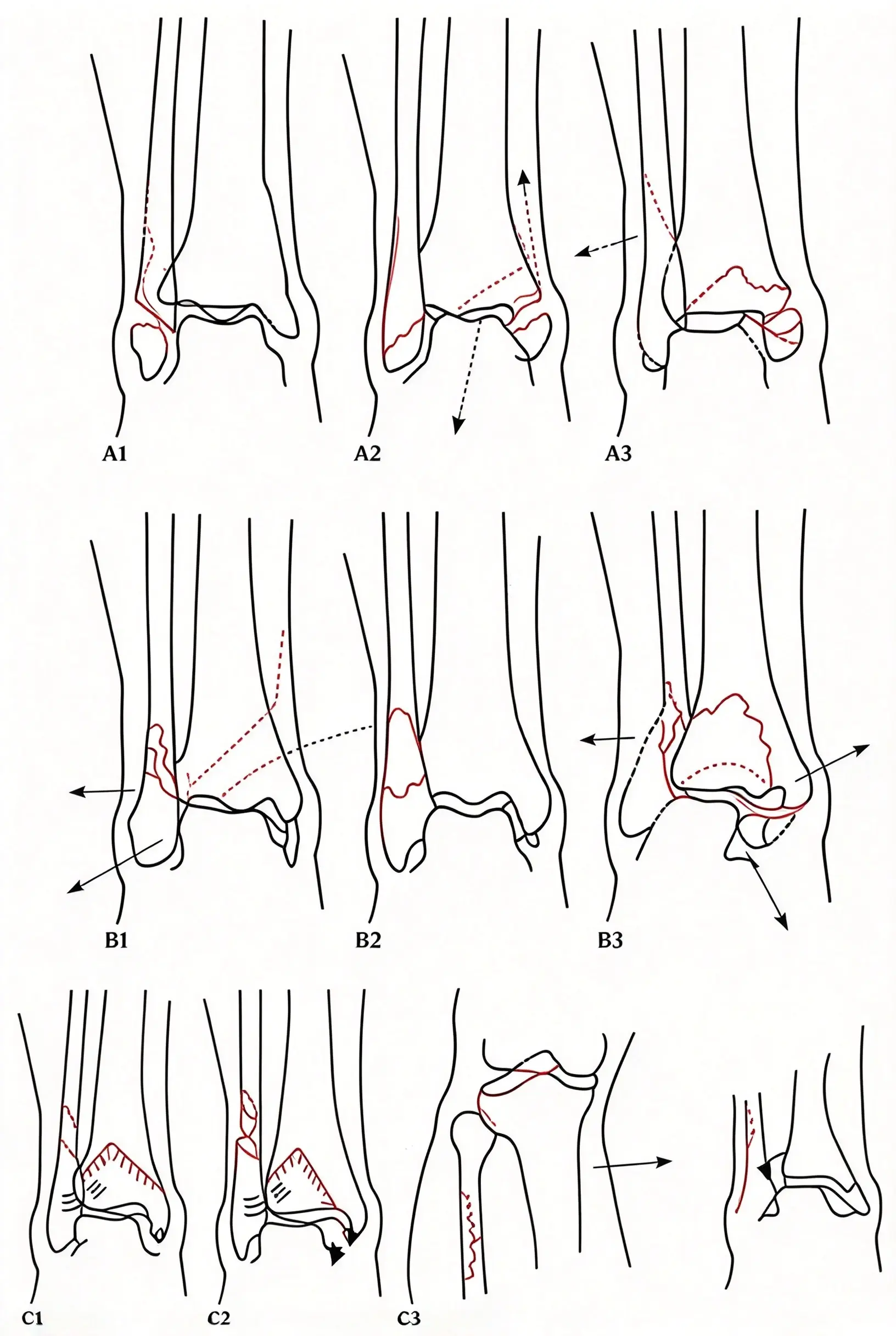
The AO/OTA classification provides a more detailed description of fracture location, morphology, and associated injuries. It is widely used in orthopedic trauma documentation, research, and clinical communication.
Ankle fractures are assigned the anatomical code 44 and are broadly divided into three groups:
44A: Infrasyndesmotic fibular fractures.
44B: Transsyndesmotic fibular fractures.
44C: Suprasyndesmotic fibular fractures.
These groups correspond broadly to the Danis-Weber classification.
Further subdivisions describe additional fracture characteristics, including the involvement of the medial and posterior structures, fracture complexity, and associated injuries.
The AO/OTA classification is particularly useful for standardized fracture documentation and clinical research. For complex trimalleolar fractures, CT imaging can provide additional information about posterior malleolar morphology and articular involvement that may not be fully appreciated on plain radiographs.
It is further subdivided into types A, B, and C with reference to the Danis-Weber classification. The third new category is further subdivided according to the degree of fracture comminution.
The posterior malleolus is an important component of ankle joint stability. Its involvement may affect the articular surface, posterior syndesmotic attachment, and congruency of the ankle mortise.
Not every posterior malleolus fracture requires surgical fixation. The decision should be based on the overall injury pattern rather than fragment size alone.
The posterior malleolus contributes to:
The posterior articular surface of the ankle joint.
Stability of the distal tibiofibular syndesmosis.
Maintenance of the ankle mortise.
Attachment of the posterior inferior tibiofibular ligament.
A displaced posterior malleolar fragment may contribute to joint incongruity, posterior talar subluxation, and syndesmotic instability.
Therefore, accurate assessment of the posterior malleolus is essential when planning treatment for a trimalleolar fracture.
Historically, surgeons often considered posterior malleolar fixation when the fracture fragment involved more than approximately 25% of the tibial plafond articular surface or when displacement exceeded 2 mm.
These measurements remain useful historical reference points, but they should not be treated as universal indications.
Current decision-making places greater emphasis on several factors:
1. Fragment size and morphology
The size, shape, and location of the posterior fragment should be evaluated. Some smaller fragments may have important implications for syndesmotic stability.
2. Articular displacement and joint congruity
Residual displacement, articular step-off, and impaction can affect ankle joint mechanics and may influence the need for fixation.
3. Syndesmotic stability
Because the posterior inferior tibiofibular ligament attaches to the posterior malleolus, restoring the posterior fragment may help restore the posterior syndesmotic attachment and improve ankle stability.
4. Associated injuries
The presence of fibular fractures, medial malleolar fractures, deltoid ligament injuries, and ankle dislocation should be considered together.
Standard ankle radiographs, including anteroposterior, mortise, and lateral views, are used for initial assessment.
However, computed tomography (CT) is particularly useful in complex posterior malleolar fractures because it can help determine:
Fragment size and configuration.
Involvement of the fibular incisura.
Articular impaction and comminution.
The presence of intercalary fragments.
The relationship between the posterior fragment and the syndesmosis.
CT-based assessment can help surgeons select an appropriate surgical approach and fixation strategy.
When fixation is indicated, the method depends on fracture morphology, fragment size, displacement, bone quality, and surgical approach.
Common techniques include:
Posterior-to-anterior screw fixation
Screws may be inserted from posterior to anterior to stabilize a suitable posterior fragment. This technique is generally performed through an appropriate posterior surgical approach when direct visualization is required.
Anterior-to-posterior screw fixation
In selected fracture patterns, screws can be inserted from anterior to posterior. This may be suitable for certain fragments, although the direction of fixation and ability to achieve adequate reduction must be considered.
Posterior buttress plate fixation
A posterior plate can provide buttress support for fragments that are prone to displacement, particularly when the fracture morphology requires direct posterior stabilization.
The objective of posterior malleolar fixation is to restore the articular surface, improve ankle stability, and address associated syndesmotic injury when appropriate. Screw fixation is one option, but plate fixation may be preferable for certain fracture configurations.
Treatment selection depends on fracture displacement, ankle stability, articular congruity, soft-tissue condition, patient factors, and associated injuries.
Both nonoperative and operative treatment may be appropriate in selected circumstances. However, displaced or unstable trimalleolar fractures frequently require surgical management.
Nonoperative treatment may be considered in carefully selected patients, such as those with:
Stable, minimally displaced fractures.
A congruent ankle mortise.
No clinically significant talar shift.
Medical conditions that make surgery excessively risky.
Other circumstances in which operative treatment is not appropriate.
Treatment may involve immobilization with a cast or removable boot, followed by regular clinical and radiographic monitoring.
A displaced or unstable trimalleolar fracture is generally not suitable for routine conservative treatment because maintaining anatomical alignment can be difficult.
Potential complications of inadequately reduced or unstable fractures include:
Persistent ankle mortise incongruity.
Malunion or nonunion.
Chronic ankle instability.
Restricted ankle range of motion.
Post-traumatic osteoarthritis.
Persistent pain and impaired weight-bearing function.
The key objective of nonoperative management is to maintain a stable, well-aligned ankle joint throughout fracture healing.
Surgical treatment is commonly recommended for displaced or unstable trimalleolar fractures, particularly when the ankle mortise is incongruent or the talus is subluxated.
The primary goals of surgery are to:
Restore the normal anatomy of the ankle joint.
Re-establish a congruent ankle mortise.
Restore fibular length and rotation.
Stabilize the medial and lateral malleoli.
Assess and address posterior malleolar involvement.
Restore syndesmotic stability when necessary.
Allow progressive rehabilitation and functional recovery.
Lateral malleolus fixation
The distal fibula is commonly stabilized using plates and screws. Depending on fracture morphology and bone quality, surgeons may select an anatomical distal fibular plate, locking plate, or other suitable fixation construct.
Medial malleolus fixation
Medial malleolar fractures may be treated with cannulated screws, partially threaded screws, tension band constructs, or plates, depending on fragment size, fracture configuration, and bone quality.
Posterior malleolus fixation
Posterior fragments may be treated with screws or posterior buttress plates when fixation is indicated. The decision should consider fragment morphology, articular reduction, and syndesmotic stability.
Syndesmotic stabilization
If instability persists after fracture reduction and fixation, additional syndesmotic stabilization may be required. Options include syndesmotic screws or suture-button devices, depending on the injury pattern and surgeon preference.
The ankle joint is a weight-bearing joint that depends on precise alignment between the tibia, fibula, and talus.
Even relatively small residual abnormalities in joint congruity or fibular position can alter load distribution across the ankle joint.
Anatomical reduction and stable fixation aim to restore joint mechanics and reduce the risk of long-term complications. Nevertheless, the final outcome also depends on cartilage damage at the time of injury, soft-tissue condition, rehabilitation, and patient-specific factors.
Rehabilitation is an important part of recovery after trimalleolar fracture fixation.
The rehabilitation plan should be individualized according to fracture stability, fixation quality, soft-tissue healing, bone quality, associated injuries, and the surgeon's postoperative instructions.
For patients with stable fixation and appropriate wound healing, controlled early rehabilitation may help reduce stiffness and maintain muscle function.
The initial goals are to control swelling, protect the surgical repair, and maintain mobility in the surrounding joints.
1. Elevation of the affected limb
Elevating the injured leg can help reduce postoperative swelling. The foot should be positioned above the level of the heart when practical, particularly during the early recovery period.
2. Controlled ankle range-of-motion exercises
When permitted by the surgeon, patients may begin gentle active or assisted ankle dorsiflexion and plantarflexion within a comfortable range.
Early movement may help reduce joint stiffness and maintain mobility.
In the initial phase, inversion and eversion exercises may be restricted depending on the fracture pattern and fixation construct.
3. Hip and knee mobility exercises
Movement of the adjacent joints helps maintain general lower-limb function.
Examples include:
Hip and knee flexion and extension.
Straight-leg raises to activate the quadriceps.
Hip abduction exercises to strengthen the gluteus medius.
Hip extension exercises to activate the gluteus maximus.
4. Early mobilization without weight-bearing
When medically appropriate, patients may begin transfers and walking with crutches or a walker while keeping the operated ankle non-weight-bearing.
Early mobilization can help reduce complications associated with prolonged immobility. However, weight-bearing restrictions must be followed carefully.
5. Cold therapy
Cold therapy may help reduce pain and swelling during the early postoperative period.
It should be applied with appropriate skin protection and according to clinical advice. Patients should avoid direct prolonged contact between ice and the skin.
As fracture healing progresses, rehabilitation may be advanced according to clinical and radiographic assessment.
Typical components include:
Gradual improvement in ankle dorsiflexion and plantarflexion.
Progressive strengthening of the calf and surrounding lower-limb muscles.
Balance and proprioception exercises.
Gait training.
Gradual progression of weight-bearing when authorized.
Functional exercises to improve walking and daily activities.
The timing of each stage varies. There is no single rehabilitation schedule suitable for every trimalleolar fracture.
Several precautions should be observed during rehabilitation:
1. Prioritize active muscle training when appropriate
Active exercises help patients regain muscle control and strength. They should be introduced according to the stage of healing and the surgeon's instructions.
2. Avoid forceful passive movement
Early passive exercises should be gentle and controlled. Excessive force may place unnecessary stress on the healing fracture or fixation construct.
3. Coordinate rehabilitation with pain management
Appropriate pain control can help patients participate in rehabilitation. Physical therapy and medication may be used together when clinically indicated.
4. Progress gradually
Exercise intensity, range of motion, and weight-bearing should increase progressively. Sudden increases in loading may compromise recovery.
5. Follow weight-bearing restrictions
Patients should not begin full weight-bearing simply because pain has decreased. Radiographic healing, fracture stability, and the surgeon's assessment should guide progression.
Despite appropriate treatment, trimalleolar fractures can be associated with complications.
Common concerns include:
Post-traumatic osteoarthritis: Cartilage damage and residual joint incongruity may contribute to degenerative changes.
Malunion: The fracture heals in an abnormal position, potentially affecting ankle alignment and function.
Nonunion: One or more fracture fragments fail to unite.
Joint stiffness: Prolonged immobilization and soft-tissue injury may restrict ankle movement.
Wound complications: Swelling, skin damage, and surgical wound problems may delay recovery.
Infection: Surgical site infection is a potential complication of operative treatment.
Implant-related problems: Screws or plates may cause irritation or require further evaluation.
Chronic instability: Inadequate restoration of the ankle mortise or syndesmotic stability may lead to persistent instability.
Early diagnosis, appropriate fracture reduction, careful fixation, soft-tissue management, and structured rehabilitation all contribute to improving outcomes.
A bimalleolar fracture involves the medial and lateral malleoli. A trimalleolar fracture involves all three malleoli: medial, lateral, and posterior. Trimalleolar fractures may be associated with greater joint instability and more complex articular involvement.
No. Treatment depends on fracture displacement, ankle stability, joint alignment, patient health, and other clinical factors. However, displaced or unstable trimalleolar fractures commonly require operative fixation to restore ankle alignment and stability.
No. Posterior malleolar fixation is not determined by fragment size alone. Surgeons also consider displacement, articular step-off, fracture morphology, syndesmotic stability, and the overall ankle injury pattern.
Diagnosis usually involves physical examination and ankle radiographs. CT scanning may be recommended for complex fractures, especially when detailed evaluation of the posterior malleolus is needed for surgical planning.
Recovery varies according to fracture severity, soft-tissue injury, fixation stability, bone healing, and rehabilitation progress. Some patients require several months to regain functional mobility, while swelling, stiffness, and strength deficits may persist longer.
The timing of weight-bearing depends on the fracture pattern, fixation construct, bone quality, and postoperative assessment. Some patients may begin earlier protected weight-bearing, while others require a longer non-weight-bearing period. The treating surgeon should determine the appropriate schedule.
A trimalleolar ankle fracture is a complex injury involving the medial, lateral, and posterior malleoli. Because these structures contribute to ankle joint congruity and stability, accurate assessment of the fracture pattern is essential.
The Lauge-Hansen, Danis-Weber, and AO/OTA classification systems provide different ways to describe ankle fracture morphology and injury mechanisms. Posterior malleolar involvement requires particular attention because it can affect the articular surface and syndesmotic stability.
For displaced or unstable fractures, surgical treatment aims to restore anatomical alignment and provide sufficient stability for fracture healing and progressive rehabilitation. The decision to fix the posterior malleolus should be based on fragment morphology, articular congruity, and ankle stability rather than a single size threshold.
With appropriate treatment, careful postoperative monitoring, and individualized rehabilitation, many patients can regain useful ankle function and return to daily activities.


