
Views: 0 Author: Site Editor Publish Time: 2026-10-26 Origin: Site
Introduction
Clavicle fractures are among the most common fractures encountered in orthopedic trauma practice. Although many clavicle fractures can be managed without surgery, displaced fractures, substantial shortening, comminution, open injuries, and selected cases with a high risk of nonunion may require operative fixation.
The two commonly used surgical options for midshaft clavicle fractures are plate fixation and intramedullary fixation. Plate fixation provides direct control of fracture alignment and can accommodate complex fracture patterns. Intramedullary fixation uses an implant placed within the medullary canal and may reduce the size of the surgical incision and the prominence of external hardware.
Neither technique is universally superior. Fracture morphology, patient characteristics, soft-tissue condition, implant design, and the surgeon's experience all influence treatment selection.
This article reviews surgical decision-making, plate selection and positioning, neurovascular protection, minimally invasive techniques, intramedullary nail indications, and common complications. It is intended for professional education and should be interpreted alongside current clinical guidance and the instructions for use of the selected implant system.
The first decision is not which implant to use, but whether surgery is appropriate.
Many clavicle fractures heal with nonoperative treatment. However, in adults with displaced midshaft fractures, operative treatment is associated with higher union rates and better early patient-reported outcomes in comparison with nonoperative treatment. Long-term functional outcomes may be similar for some patients, so the decision should balance the expected benefits against surgical risks and the possibility of subsequent implant removal.
Clinical assessment should consider:
Fracture displacement and shortening.
Comminution and the stability of the fracture pattern.
Skin condition and the risk of skin compromise.
Open injury or associated neurovascular injury.
Patient age, activity level, occupation, and functional requirements.
Smoking status and other factors that may impair bone healing.
Patient preferences and willingness to accept surgical risks or implant-related symptoms.
A displaced fracture does not automatically require surgery in every patient. Conversely, a high-risk fracture in a patient with additional risk factors may justify early operative discussion.
Smoking, substantial displacement, and comminution are important factors when estimating the risk of nonunion. Their combined presence may strengthen the case for operative treatment, but the decision should be based on the complete clinical picture.
The original source article cites a Murray-model estimate suggesting that the number needed to treat (NNT) to prevent one nonunion may be approximately 7.5 when all displaced fractures are treated surgically, compared with approximately 1.7 in a high-risk subgroup with smoking, comminution, and marked displacement.
These figures should not be treated as universal estimates. NNT depends on the underlying population, baseline nonunion risk, outcome definition, and treatment effect. Before publishing these exact numbers as clinical evidence, verify them against the original study and explain the population to which they apply.
Patients initially managed without surgery require appropriate follow-up. Persistent pain, poor functional recovery, fracture-site mobility, and absent radiographic progression may raise concern for delayed healing or nonunion.
The original article proposes a reassessment point at approximately six weeks and highlights three findings:
A QuickDASH score of approximately 40.
No bridging callus on radiographs or ultrasound.
Abnormal mobility at the fracture site on examination.
These findings may be clinically useful prompts for reassessment, but they should not be presented as a validated universal rule stating that two findings mandate immediate surgery. A single time point cannot reliably establish nonunion in every patient.
The treating clinician should interpret symptoms, physical examination, serial imaging, fracture pattern, and the patient's recovery together. When healing is uncertain, further assessment and individualized treatment planning are preferable to an automatic operative threshold.
Plate fixation is commonly used for displaced midshaft clavicle fractures, particularly when the fracture is comminuted or requires direct control of length, rotation, and alignment.
The key technical challenges extend beyond screw placement. They include choosing an appropriate implant, preserving the soft-tissue envelope, protecting the underlying neurovascular structures, and minimizing hardware-related irritation.
The original article recommends avoiding conventional 3.5 mm reconstruction plates in favor of anatomically contoured locking plates or a dual-mini-plate construct.
A more evidence-based interpretation is that plate selection should reflect fracture morphology, required mechanical stability, bone quality, plate contour, and the intended fixation strategy.
A conventional reconstruction plate can be useful in selected cases, particularly where contouring is important. However, a plate that is insufficiently stiff for the fracture pattern may be more susceptible to deformation or fatigue failure under repeated loading.
For fractures with substantial comminution, a suitably selected precontoured clavicle plate or an appropriately designed dual-plate construct may provide the required stability. The choice should be based on the implant system's mechanical characteristics and the surgeon's fixation plan rather than a universal prohibition against one plate type.
The source article reports a fatigue-failure figure of up to 53% for reconstruction plates in complex comminuted fractures. This figure requires verification against the original study before publication, because failure rates depend on plate design, fracture pattern, patient loading, and study population.
Patient positioning can affect the spatial relationship between the clavicle, plate, screws, and nearby neurovascular structures.
The beach-chair position is frequently used for clavicle fixation, but it should not be described as universally safer for every plate configuration. Cadaveric research indicates that the relationship between patient position and neurovascular risk differs between superior and anteroinferior plating. The medial screw holes deserve particular attention with either configuration.
The operative position should therefore be selected according to the planned plate location, the patient's anatomy, imaging requirements, and the surgeon's technique.
Two commonly discussed plate positions are the superior surface and the anteroinferior surface of the clavicle.
Superior plating
Potential advantages:
Provides a familiar and direct fixation surface.
Can offer strong mechanical fixation when appropriately applied.
May be suitable for a range of fracture patterns.
Potential disadvantages:
The plate may be prominent beneath the skin.
Hardware irritation may lead to discomfort or later implant removal.
Drilling and screw insertion require careful protection of the structures beneath the clavicle.
Anteroinferior plating
Potential advantages:
May reduce superior hardware prominence in selected patients.
Can provide a useful screw trajectory and fixation construct for appropriate fracture patterns.
May be advantageous when the plate contour and soft-tissue coverage suit the patient's anatomy.
Potential disadvantages:
The approach does not eliminate neurovascular risk.
Screw direction and length require careful planning.
The safest plate position depends on individual anatomy and the selected implant.
The original article favors anteroinferior plating to reduce soft-tissue irritation and improve the mechanical construct. These potential benefits should be described as technique-dependent rather than guaranteed outcomes. Published anatomical studies have found that both plate positions can place nearby structures at risk.
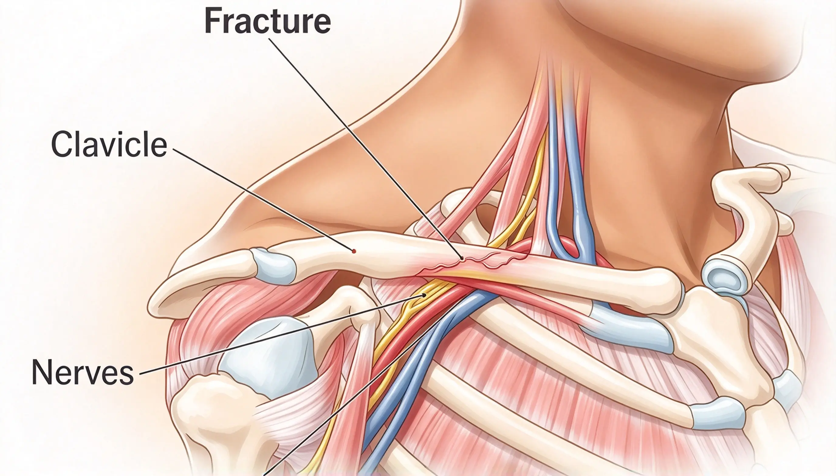
The clavicle lies close to important structures, including the subclavian vessels and brachial plexus. The risk of injury is especially important during drilling through the posterior cortex and when inserting screws into the medial and middle portions of the bone.
The subclavian vessels lie close to the posterior aspect of the medial clavicle. The exact distance varies with individual anatomy, patient position, and the direction of drilling.
The source article cites an average distance of approximately 4.77 mm, with some measurements reported as zero. Such measurements are highly dependent on the anatomical study and measurement method and should not be interpreted as a universal safe-distance estimate.
Practical safety principles include:
Plan the drilling trajectory according to the plate position and local anatomy.
Use appropriate drill guides, depth control, and protective instruments.
Avoid uncontrolled advancement of the drill beyond the far cortex.
Select screw length based on the measured bone and the implant system's recommendations.
Consider whether bicortical purchase is necessary at a particular location.
The source article suggests unicortical fixation for the most medial screw or a deliberately altered drilling direction in selected situations. This should not be applied as a universal rule. The decision between unicortical and bicortical fixation depends on the fracture, construct, bone quality, and implant instructions.
The subclavian vessels and brachial plexus remain at risk during drilling in the middle portion of the clavicle. The risk depends on the screw hole, drilling direction, plate configuration, and patient position.
Safety measures include:
Using a suitable drill stop or depth-limiting system when appropriate.
Measuring the required drilling depth rather than relying on a fixed depth for every patient.
Protecting the far cortex and adjacent soft tissues with an appropriate instrument when the operative technique permits.
Maintaining control of the drill and avoiding excessive penetration.
Confirming screw length and final implant position.
The source article mentions a drill stop set to 15–20 mm. This range should not be used as a universal setting because clavicle dimensions and implant systems differ. Depth must be determined from the patient's anatomy and the intended fixation construct.
The neurovascular structures generally become more distant from parts of the lateral clavicle, but this does not make the entire lateral region safe for unrestricted drilling.
The lateral clavicle has its own anatomical considerations, including the acromioclavicular joint, nearby soft tissues, and the fixation requirements of distal fractures. The screw trajectory should always be controlled and appropriate to the planned construct.
Key principle: No part of the clavicle should be treated as a region where uncontrolled drilling is acceptable. Protection of the far cortex and awareness of the surrounding anatomy are essential throughout the procedure.
In comminuted fractures, aggressive exposure and extensive dissection can compromise the soft-tissue attachments and blood supply of intermediate fragments.
The aim of bridge plating is to restore the overall length, alignment, and rotation of the clavicle while preserving the biological environment around the fracture. Individual fragments do not always need to be anatomically reconstructed if the fracture pattern and fixation strategy permit stable indirect reduction.
Minimally invasive plate osteosynthesis may be considered in selected fracture patterns. The technique aims to reduce unnecessary soft-tissue disruption while achieving an appropriate fixation construct.
Important principles include:
Preserving soft-tissue attachments to intermediate fragments where possible.
Avoiding excessive periosteal stripping.
Maintaining appropriate clavicle length and rotation.
Ensuring adequate fixation on both sides of the fracture.
Confirming alignment and implant position with suitable imaging.
MIPO is not automatically appropriate for every comminuted fracture. Some fracture patterns require direct visualization or a different reduction strategy to achieve acceptable alignment and stability.
The original article cites a reduction in postoperative anterior chest-region numbness from 55% to 12.5% with a minimally invasive technique. This numerical comparison should be included only after the original study and its outcome definitions have been verified.
Plate prominence, local irritation, patient preference, and other implant-related issues may lead to consideration of plate removal after fracture union.
However, removal is not risk-free. Screw holes can act as stress concentrators, and the clavicle may remain vulnerable to refracture after hardware removal.
The original article reports a refracture rate of approximately 9% within two months of plate removal. This should not be presented as a universal rate without verifying the original study and its patient population.
Before implant removal, clinicians should consider:
Whether the fracture has achieved adequate union.
The severity of symptoms attributable to the implant.
Patient-specific risks and activity requirements.
The potential benefits of removal versus continued observation.
Post-removal activity restrictions and follow-up.
Patients should understand that removal is not always necessary and that refracture remains a possible complication. After removal, activity progression should follow the treating clinician's instructions.
Intramedullary fixation uses an implant placed within the clavicular medullary canal to stabilize the fracture. Devices may include titanium elastic nails (TENs), elastic stable intramedullary nails (ESIN), or other clavicle-specific intramedullary implants.
Compared with plate fixation, intramedullary devices may require smaller incisions and may reduce the prominence of hardware in selected patients. However, they may provide less control of rotation, shortening, and complex comminution, depending on the implant design and fracture pattern.
The American Academy of Orthopaedic Surgeons (AAOS) clinical practice guideline states that intramedullary fixation and single-plate fixation can achieve similar long-term clinical outcomes for clavicle shaft fractures. Plate fixation may be preferable when the fracture is comminuted. AAOS Clinical Practice Guideline
Intramedullary fixation is generally better suited to selected simple midshaft fractures, such as transverse or short-oblique patterns, where length and alignment can be controlled with the chosen implant.
The original article identifies Robinson 2B1 fractures as the preferred indication and advises against using elastic nails for comminuted or length-unstable fractures.
This is a useful general distinction, but implant selection should not rely on the classification label alone. Fracture morphology, shortening, canal dimensions, implant design, and the surgeon's ability to obtain stable fixation all matter.
For markedly comminuted or length-unstable fractures, plate fixation is often more suitable because it can provide greater control over length, rotation, and fragment alignment.
The entry point depends on the specific intramedullary device and its approved surgical technique. The source article describes a medial entry point near the sternoclavicular joint and emphasizes avoiding an unsuitable lateral entry trajectory.
The exact entry location should be determined from the device instructions, preoperative imaging, and the patient's anatomy. A fixed distance from the sternoclavicular joint should not be applied to every implant system.
Careful preparation of the entry site and controlled insertion are important because the medial clavicle is close to the thoracic inlet and adjacent neurovascular structures.
Closed reduction may be challenging in some clavicle fractures because of shortening, fragment overlap, soft-tissue interposition, or fracture geometry.
If a controlled closed-reduction attempt is unsuccessful, the surgeon may consider a limited open approach when appropriate. A small incision can allow direct assistance with reduction while limiting unnecessary dissection.
Forceful blind advancement of the implant should be avoided. Uncontrolled passage can damage the surrounding soft tissues and, in rare cases, injure the pleura or other structures near the thoracic inlet.
A single elastic intramedullary implant may provide limited resistance to rotation or axial shortening in certain fracture patterns. Telescoping can occur when fracture stability and implant characteristics do not adequately control the fragments.
Risk reduction includes:
Selecting a suitable fracture pattern for the implant.
Confirming satisfactory reduction and alignment.
Following the device-specific fixation technique.
Providing postoperative protection appropriate to the fracture and construct.
Progressing shoulder movement and loading according to the treating clinician's protocol.
The source article recommends sling immobilization for the first three to four weeks and avoiding shoulder abduction or forward flexion beyond 90 degrees until callus develops. These restrictions should not be presented as a universal protocol. Rehabilitation depends on fracture stability, the implant, postoperative findings, and the surgeon's instructions.
Implant migration or back-out can cause discomfort and irritation near the entry site. In some cases, prominent hardware can threaten the overlying skin.
The original article recommends terminal end-caps to reduce implant migration. Whether an end-cap is appropriate depends on the specific nail system and its approved design. Some implants use end-caps or other mechanisms to improve retention; these features should be used according to the manufacturer's instructions rather than being treated as mandatory for every intramedullary device.
Postoperative follow-up should assess implant position, entry-site symptoms, skin condition, fracture alignment, and healing progression.
Intramedullary implants may also require removal because of irritation, migration, or the device's intended treatment pathway. A smaller implant does not guarantee zero refracture risk after removal.
The source article describes the refracture risk after elastic nail removal as zero. This absolute claim should not be used without strong, directly applicable evidence. Refracture risk depends on fracture union, residual bone weakness, implant design, removal technique, and subsequent activity.
| Clinical consideration | Plate fixation | Intramedullary fixation |
|---|---|---|
| Simple midshaft fracture | Commonly used; provides direct control of alignment | Suitable for selected fractures |
| Comminuted fracture | Often advantageous for restoring length and controlling fragments | May be less suitable when length or rotation is unstable |
| Rotational control | Can provide strong control when appropriately constructed | Depends on nail design and fracture pattern |
| Soft-tissue exposure | May require a larger incision and more dissection | Often uses smaller incisions |
| Hardware prominence | Can cause irritation, especially with prominent plates | Entry-site irritation or nail migration may occur |
| Neurovascular safety | Requires careful drilling and screw-length control | Requires safe entry and controlled implant passage |
| Implant removal | May be considered for symptomatic hardware; refracture is possible | May be required for irritation, migration, or device-specific reasons |
| Treatment selection | Particularly useful for complex or comminuted patterns | Best reserved for fractures that can be adequately controlled by the selected implant |
Neither option is appropriate for every fracture. The final choice should reflect the fracture pattern, patient factors, implant design, and the surgeon's assessment.
No. Surgery can improve union rates and early functional outcomes in selected adults with displaced midshaft fractures, but treatment must be individualized. Many patients can still be managed nonoperatively.
Neither is universally better. Both can provide good long-term outcomes in suitable fractures. Plates are often more appropriate for comminuted fractures, while intramedullary fixation may be suitable for selected simple fracture patterns.
No. Plate position changes the relationship between the implant, screws, and underlying neurovascular structures. Both techniques require careful planning and controlled drilling. The safest configuration depends on anatomy, patient position, and the implant system.
The subclavian vessels and brachial plexus lie close to the posterior surface of the clavicle. Uncontrolled drill penetration or an excessively long screw can injure these structures. Depth control, careful trajectory planning, and protection of the far cortex are important.
Plate removal may be considered for persistent hardware-related symptoms after adequate fracture healing. It is not necessary in every patient, and removal carries risks, including refracture. The decision should be made with the treating surgeon.
They may be unsuitable when the fracture is highly comminuted or cannot maintain length and rotation with the selected device. Plate fixation often offers better control in these situations.
No. End-cap requirements and implant-retention mechanisms vary by device. The appropriate technique is the one specified by the implant manufacturer and selected for the patient's fracture.
Clavicle fracture fixation requires more than choosing between a plate and an intramedullary nail. Successful treatment begins with appropriate patient selection and continues through fracture reduction, implant positioning, protection of the underlying neurovascular structures, and individualized postoperative rehabilitation.
Plate fixation is often advantageous for comminuted or length-unstable fractures because it provides direct control of alignment. Intramedullary fixation can be a useful minimally invasive option for selected simple midshaft fractures, provided the implant can adequately control the fracture pattern.
The most important safety principles are to avoid treating numerical thresholds as universal rules, maintain control during drilling and implant insertion, preserve fracture biology where possible, and follow the instructions for the selected fixation system.
For orthopedic professionals evaluating clavicle fixation systems, implant design, mechanical performance, anatomical fit, compatible instruments, and clear surgical instructions should all be considered when selecting a solution.
American Academy of Orthopaedic Surgeons (AAOS). Treatment of Clavicle Fractures: Clinical Practice Guideline. View the guideline
Lo EY, Eastman J, Tseng S, Lee MA, Yoo BJ. Neurovascular risks of anteroinferior clavicular plating. Orthopedics. 2010;33(1):21. PubMed
Patient Position Is Related to the Risk of Neurovascular Injury in Clavicular Plating: A Cadaveric Study. PubMed
Surgical interventions for treating acute fractures or non-union of the middle third of the clavicle. Cochrane Database of Systematic Reviews. Full review
Clinical disclaimer: This article is intended for professional education and is not a substitute for formal surgical training, patient-specific assessment, current clinical guidance, or the instructions for use supplied with the relevant implant system.


