English


Views: 0 Author: Site Editor Publish Time: 2026-10-28 Origin: Site
Introduction
Proximal humerus fractures (PHFs) are challenging injuries, particularly in older adults with osteoporosis. The complex anatomy of the shoulder, vulnerable blood supply to the humeral head, and deforming forces generated by the rotator cuff can make fracture reduction and stable fixation difficult. Complications may include varus collapse, screw penetration into the glenohumeral joint, fixation failure, avascular necrosis (AVN), and loss of shoulder function.
Successful treatment requires more than selecting an implant. Surgeons must understand the fracture pattern, preserve soft-tissue attachments, restore medial column support, and choose between open reduction and internal fixation (ORIF) and arthroplasty according to the patient's age, bone quality, fracture morphology, and functional needs.
This article reviews 20 practical tips and common pitfalls in proximal humerus fracture surgery, focusing on deltopectoral exposure, PHILOS locking plate fixation, osteoporotic bone augmentation, reverse total shoulder arthroplasty (RTSA), humeral head viability, and intraoperative screw-length assessment.
The deltopectoral approach is commonly used for proximal humerus fracture fixation because it provides access to the anterior shoulder while preserving the deltoid muscle.
During exposure, identify the cephalic vein early and protect it throughout the procedure. The vein is commonly mobilized with the deltoid toward the lateral side, although the direction of mobilization should be adapted to the anatomy, venous branches, and surgeon's preferred technique. The pectoralis major and conjoint tendon are managed as necessary to establish a safe working corridor.
Avoid excessive traction or unnecessary ligation of venous branches. Gentle tissue handling helps reduce bleeding and preserves the soft-tissue envelope.
The axillary nerve is vulnerable during proximal humerus surgery, particularly when retractors are placed around the inferior capsule or proximal humerus.
Maintain awareness of the nerve's course and avoid forceful or prolonged deep retraction. Reassess retractor position whenever exposure changes, especially during manipulation of the humeral shaft or inferior fracture fragments.
A traction-based assessment, sometimes informally described as a “tug test,” should not be regarded as a validated method for excluding axillary nerve compression. Direct anatomical awareness, careful retractor placement, and repeated reassessment are more reliable safety principles.
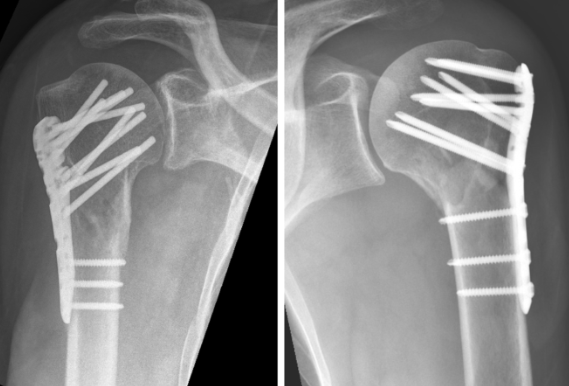
PHILOS-type proximal humerus locking plates provide angular-stable fixation and are widely used for selected displaced fractures. However, locking screws alone cannot compensate for poor reduction, deficient medial support, or inadequate bone quality.
Medial column support is one of the most important factors in maintaining reduction after proximal humerus fracture fixation.
Medial calcar comminution, residual varus, and loss of the medial hinge can increase the risk of varus collapse and fixation failure. Whenever possible, restore the medial cortical buttress and achieve a stable reduction before definitive plate fixation.
A plate may appear well positioned on an anteroposterior image while the medial column remains inadequately supported. Evaluate the reduction in multiple fluoroscopic views.
Inferomedial calcar screws are designed to reinforce the humeral head near the medial column, helping resist varus deformation in appropriately reduced fractures.
Their effectiveness depends on the fracture configuration, reduction quality, screw trajectory, and bone stock. Use the dedicated plate holes and follow the implant system's recommended trajectories. Confirm screw position fluoroscopically and avoid penetration into the joint.
A small trajectory error may substantially reduce the intended mechanical support. However, a specific displacement threshold, such as an 8 mm deviation, should not be treated as a universal point at which construct stiffness suddenly collapses; the effect depends on the implant geometry and fracture model.
A commonly described positioning target for some proximal humerus plates is approximately 5–8 mm distal to the apex of the greater tuberosity. The plate is also often positioned slightly posterior to the bicipital groove to reduce the risk of impingement and avoid injury to adjacent structures.
These measurements are reference points, not rules that apply to every plate design.
Different plate systems have different proximal contours, screw trajectories, and recommended positioning. Some designs require a more distal placement than others. Therefore:
Review the manufacturer's surgical technique before surgery.
Confirm that the plate does not impinge on the acromion during shoulder movement.
Check the relationship between the plate, greater tuberosity, and bicipital groove.
Confirm that proximal screws follow the intended trajectories without penetrating the joint.
The key principle is to follow the instructions for the specific implant rather than transferring a positioning measurement from another plate system.
Plate position relative to the bicipital groove influences the relationship between the implant, the long head of the biceps tendon, and the surrounding soft tissues.
The plate is commonly placed slightly posterior to the groove, but the exact offset depends on implant design and patient anatomy. Avoid excessive anterior positioning that may compromise the tendon or surrounding structures, and avoid a position that creates subacromial impingement.
The anterior circumflex humeral artery and its branches contribute to the blood supply of the humeral head. Although preserving vascular structures is important, plate position alone cannot guarantee preservation of humeral head perfusion.
In markedly comminuted fractures, the original humeral height may be difficult to reconstruct from the fracture fragments alone.
The superior border of the pectoralis major tendon insertion can provide an additional anatomical reference for estimating humeral shaft and head relationships. A distance of approximately 5.6 cm has been described in anatomical discussions, but it should not be applied as a fixed measurement in every patient.
Body size, individual anatomy, fracture displacement, and the condition of the soft tissues can affect the reliability of this landmark. Combine it with the contralateral shoulder when available, preoperative imaging, fluoroscopy, and other anatomical references.
Osteoporotic cancellous bone may provide inadequate purchase for conventional locking screws, increasing the risk of screw loosening, penetration, or loss of reduction.
Polymethylmethacrylate (PMMA) augmentation has been investigated as a method of improving screw anchorage in selected proximal humerus fractures. Some studies report improved fixation strength or fewer mechanical complications, but the magnitude of benefit varies with technique, fracture pattern, and study design.
Cement augmentation is not appropriate for every fracture. Potential concerns include cement leakage, thermal effects, difficulty during revision, and complications associated with intra-articular or extraosseous cement.
Use only a validated augmentation technique compatible with the chosen implant system. Do not interpret a reported odds ratio from an individual meta-analysis as a guaranteed reduction in risk for every patient.
A fibular strut allograft can provide structural support in selected comminuted proximal humerus fractures, particularly when the medial cortex is compromised and the humeral head requires additional support against varus collapse.
The graft is generally placed within the humeral canal to support the humeral head and restore medial column mechanics. Its position should be determined by the fracture configuration and the intended load-bearing role.
The graft should be aligned appropriately with the humeral shaft, with attention to fracture reduction, available canal space, and the planned screw trajectories. A fixed insertion angle of 0 degrees should not be considered a universal requirement.
Fibular strut augmentation can improve structural support in selected cases, but it does not guarantee superior long-term shoulder function.
A systematic review of augmentation techniques describes potential benefits for maintaining reduction, while comparative clinical evidence remains variable. Some studies have not demonstrated a clear functional advantage over other fixation strategies.
The possibility of future revision is also important. If fixation fails and conversion to shoulder arthroplasty becomes necessary, an incorporated intramedullary graft may complicate reconstruction.
Consider graft augmentation when the mechanical problem justifies it, rather than using it routinely in every osteoporotic fracture.
Reverse total shoulder arthroplasty (RTSA) has an important role in selected older patients with complex proximal humerus fractures. Unlike conventional anatomical shoulder arthroplasty, RTSA relies more heavily on the deltoid muscle for elevation, making it useful in some patients whose rotator cuff function is compromised.
However, it is not automatically the preferred treatment for every three- or four-part fracture.
The DelPhi randomized controlled trial compared RTSA with ORIF using an angular-stable plate in patients aged 65–85 years with displaced OTA/AO type 11-B2 and 11-C2 fractures.
At two years, the mean Constant score was 68.0 in the RTSA group and 54.6 in the ORIF group. In the type-C2 subgroup, the scores were 69.3 and 50.6, respectively, favoring RTSA. In the type-B2 subgroup, the difference was not statistically significant at that follow-up point. Read the DelPhi trial.
These results support RTSA as an important option for selected elderly patients with complex displaced fractures. They do not establish a universal rule that every three-part fracture should undergo ORIF or every four-part fracture should undergo RTSA.
The decision should also account for bone quality, reconstructability of the humeral head, tuberosity displacement, rotator cuff condition, pre-injury function, medical comorbidities, patient goals, and the surgeon's experience.
Chronological age alone is insufficient to determine whether a patient should receive fixation or arthroplasty.
A relatively active older patient with good bone quality and a reconstructable fracture may be a candidate for fixation. Conversely, severe osteoporosis, an unreconstructable head-split fracture, major articular destruction, or a high risk of fixation failure may favor arthroplasty.
The treatment decision should be individualized rather than based on an age cutoff or fracture-part count alone.
Although RTSA can provide elevation through the deltoid, healing of the greater and lesser tuberosities remains important for restoring external and internal rotation after fracture arthroplasty.
Reduce the tuberosities anatomically where possible and use a secure suture construct appropriate for the implant and bone quality. High-strength sutures, including suitable No. 2 nonabsorbable or high-strength suture materials, may be used according to the operative technique.
A tension-band configuration or a figure-of-eight construct may help stabilize the tuberosities. The exact pattern should be adapted to the fracture and implant rather than regarded as a mandatory named technique.
Stem-mediated cerclage techniques can help secure the tuberosities around a fracture-specific humeral stem. Sutures are passed through designated stem features or around the stem, then incorporated into the tuberosity fixation construct according to the manufacturer's technique.
The aim is to resist fragment displacement and promote healing while maintaining appropriate tension. Avoid excessive tension that could distort the tuberosities or compromise their blood supply.
Cancellous bone from the excised humeral head may be used as autograft around the tuberosities when suitable. Grafting can fill defects and support healing, but it cannot compensate for inadequate reduction or unstable fixation.
The Hertel criteria use fracture characteristics such as a short metaphyseal extension, disruption of the medial hinge, and certain complex fracture patterns to estimate the risk of humeral head ischemia.
These criteria can help identify higher-risk injuries, but they do not establish with certainty whether the humeral head will develop avascular necrosis.
A 2026 retrospective study of older patients with intracapsular proximal humerus fractures reported a high negative predictive value but a much lower positive predictive value for its Hertel-based risk classification. The findings reinforce that a high-risk classification should not be equated with inevitable osteonecrosis. Read the study.
Avascular necrosis is influenced by fracture morphology, vascular injury, biological healing, reduction, and other patient-specific factors. Imaging-based risk assessment should inform the overall treatment plan rather than determine it in isolation.
When head preservation is being considered, minimize unnecessary stripping of the periosteum and soft tissues attached to the fracture fragments.
Careful handling may help preserve residual vascular connections and the biological environment needed for healing. However, no reduction maneuver or soft-tissue-preserving technique can guarantee that the humeral head will remain viable.
In younger patients, the potential value of preserving the native joint should be balanced against fracture reconstructability, articular damage, and the likelihood of obtaining stable fixation.
Screw penetration into the glenohumeral joint is a major complication of proximal humerus locking plate fixation. It can occur during initial fixation or later when the humeral head collapses around screws that were initially extra-articular.
Measure each screw trajectory carefully and verify screw length using the implant system's recommended technique.
In some situations, a surgeon may choose a screw slightly shorter than the measured depth to provide a margin against measurement error and subsequent collapse. A proposed 2–4 mm reduction is not a universal rule, however. Excessively short screws may provide inadequate purchase, particularly in osteoporotic bone.
The appropriate length depends on the screw trajectory, humeral head dimensions, fracture reduction, bone quality, and the implant's design. Confirm that the selected screw provides adequate fixation without breaching the articular surface.
Static anteroposterior and scapular Y views may fail to reveal every instance of screw penetration.
Use multiple fluoroscopic views and, when appropriate, controlled internal and external rotation of the arm to assess the relationship between screw tips and the humeral head's subchondral contour.
A dynamic fluoroscopic sweep can help reveal screws that approach or penetrate the joint surface in certain positions. If penetration is suspected, reassess the relevant trajectory and exchange the screw for an appropriately shorter one when indicated.
Do not rely on a single view or assume that a screw is safe simply because its tip appears to follow the contour of the humeral head in one projection.
Before completing fixation, review the overall construct rather than checking each screw independently.
Confirm:
Humeral head alignment and restoration of appropriate head-shaft relationships.
Medial column support and the absence of significant residual varus.
Greater and lesser tuberosity reduction where relevant.
Plate position relative to the greater tuberosity and bicipital groove.
Screw lengths and trajectories in multiple fluoroscopic projections.
Absence of obvious subacromial impingement or intra-articular penetration.
Repeat the assessment after definitive fixation if tightening or fragment manipulation could have changed the reduction.
A successful operation should consider not only immediate fixation but also the possibility of delayed collapse, screw penetration, nonunion, or avascular necrosis.
Patients with poor bone quality, severe medial comminution, residual varus, or complex articular injuries may require closer clinical and radiographic follow-up. Rehabilitation should be individualized to the fracture pattern, fixation stability, soft-tissue repair, and surgeon's protocol.
When choosing between fixation, augmentation, and arthroplasty, consider the consequences of each option if the initial strategy fails. The best construct is not necessarily the most elaborate one; it is the one that addresses the patient's specific mechanical and biological risks.
| Clinical factor | PHILOS plate fixation | Reverse total shoulder arthroplasty |
|---|---|---|
| Reconstructable humeral head | May be appropriate | May be considered in selected cases |
| Severe osteoporosis | Higher risk of fixation failure; augmentation may be considered | May be favored when reliable fixation is unlikely |
| Medial column comminution | Requires restoration or augmentation of support | May be considered based on overall fracture complexity |
| Complex displaced fracture in an older adult | Depends on reconstructability and bone quality | Supported by evidence for selected patients |
| Tuberosity healing | Important for cuff function and shoulder mechanics | Important for rotation and overall function |
| Long-term considerations | Risk of collapse, screw penetration, and osteonecrosis | Implant-related complications and tuberosity nonhealing remain concerns |
No single row should be used as an independent treatment rule. Final selection requires a complete assessment of fracture morphology, patient factors, and the expected benefits and risks of each option.
Achieving a stable reduction with adequate medial column support is fundamental. Plate position, calcar screw placement, and careful assessment of proximal screw trajectories all contribute to construct stability.
Approximately 5–8 mm below the greater tuberosity is a commonly described reference for some plate systems. The correct position varies with implant design and patient anatomy. Surgeons should follow the specific manufacturer's technique and confirm that the plate does not cause impingement.
No. Hertel criteria estimate ischemic risk but cannot reliably predict every patient's outcome. A high-risk pattern should prompt careful consideration of fracture morphology, vascular preservation, reconstructability, and follow-up rather than automatically ruling out head preservation.
It may be considered in selected fractures with medial column deficiency, particularly when additional structural support is needed to resist varus collapse. Evidence supports potential mechanical benefits, but functional superiority is not guaranteed.
No. The DelPhi trial supports RTSA for selected older patients with displaced fractures, with particularly favorable results in the studied C2 subgroup. Treatment should still be individualized according to fracture pattern, bone quality, functional expectations, and patient health.
Careful screw measurement, appropriate screw-length selection, multiple fluoroscopic views, and dynamic assessment when indicated can reduce the risk. Postoperative surveillance is also important because secondary penetration may occur after humeral head collapse.
Proximal humerus fracture surgery requires a balance between mechanical stability, biological preservation, and long-term shoulder function. The most important principles include protecting the soft tissues during exposure, restoring medial column support, positioning the PHILOS plate according to its specific design, using augmentation selectively, and assessing screw penetration in multiple views.
For complex fractures in older adults, RTSA is an important alternative to fixation, but treatment decisions should not rely on age or fracture classification alone. Likewise, Hertel criteria should be used as risk-assessment tools rather than definitive predictions of humeral head survival.
A systematic approach to reduction, implant positioning, intraoperative imaging, and postoperative follow-up can help surgeons anticipate common pitfalls and make better-informed treatment decisions.
Fraser AN, et al. Reverse Shoulder Arthroplasty Is Superior to Plate Fixation at 2 Years for Displaced Proximal Humeral Fractures in the Elderly: A Multicenter Randomized Controlled Trial. Journal of Bone and Joint Surgery. 2020. PubMed
Can Hertel Criteria Reliably Predict Avascular Necrosis After Intracapsular Proximal Humerus Fractures in the Elderly? A Retrospective Analysis. 2026. PubMed
Bone Grafting Augmentation Choices in Complex Proximal Humerus Fractures: A Systematic Review. Full text
Cement-Augmented Screw Fixation with PHILOS Plating for Osteoporotic Proximal Humeral Fractures: An Observation of Mid- and Long-Term Curative Efficacy. Orthopaedic Surgery. 2023. Publisher


