
Views: 0 Author: Site Editor Publish Time: 2026-10-21 Origin: Site
Introduction
Distal ulna fractures are frequently associated with distal radius fractures (DRFs) and commonly occur after a fall onto an outstretched hand. Although many distal ulna fractures can be managed without direct fixation, displaced or unstable fractures may require open reduction and internal fixation (ORIF) to restore anatomical alignment, improve distal radioulnar joint (DRUJ) stability, and facilitate early rehabilitation.
The distal ulna includes the ulnar styloid, ulnar head, and distal ulnar metaphysis and neck. The term distal ulnar neck fracture generally refers to a fracture in the region immediately distal to the ulnar head; the exact anatomical definition should follow the classification used in the relevant clinical source.
Treatment remains controversial, particularly when a distal ulna fracture accompanies a distal radius fracture. Fixation of the radius may restore DRUJ stability in some cases, reducing the need for a separate ulnar procedure. However, persistent instability, articular incongruity, substantial displacement, or an unstable distal ulna fracture may justify direct fixation.
This article reviews the anatomy, indications and contraindications for fixation, surgical exposure, plate positioning, fixation steps, intraoperative assessment, and strategies for preventing complications associated with distal ulna fracture ORIF.
Understanding the anatomy of the distal ulna is essential for choosing the surgical approach, selecting an appropriate plate, and avoiding injury to adjacent tendons, nerves, and joints.
The distal ulna can be divided into several clinically relevant regions:
Ulnar styloid: The distal projection of the ulna, which provides attachment for important soft-tissue structures associated with DRUJ stability.
Ulnar head: The distal articular portion of the ulna that articulates with the sigmoid notch of the radius.
Distal ulnar neck and metaphysis: The transition between the ulnar head and the distal shaft.
Distal ulnar shaft: The more proximal portion of the ulna, where fracture morphology and fixation requirements may differ from those of the ulnar head or neck.
The DRUJ permits forearm pronation and supination. Its stability depends on the congruity of the articular surfaces, the surrounding soft tissues, and the integrity of the stabilizing structures, including the triangular fibrocartilage complex (TFCC).
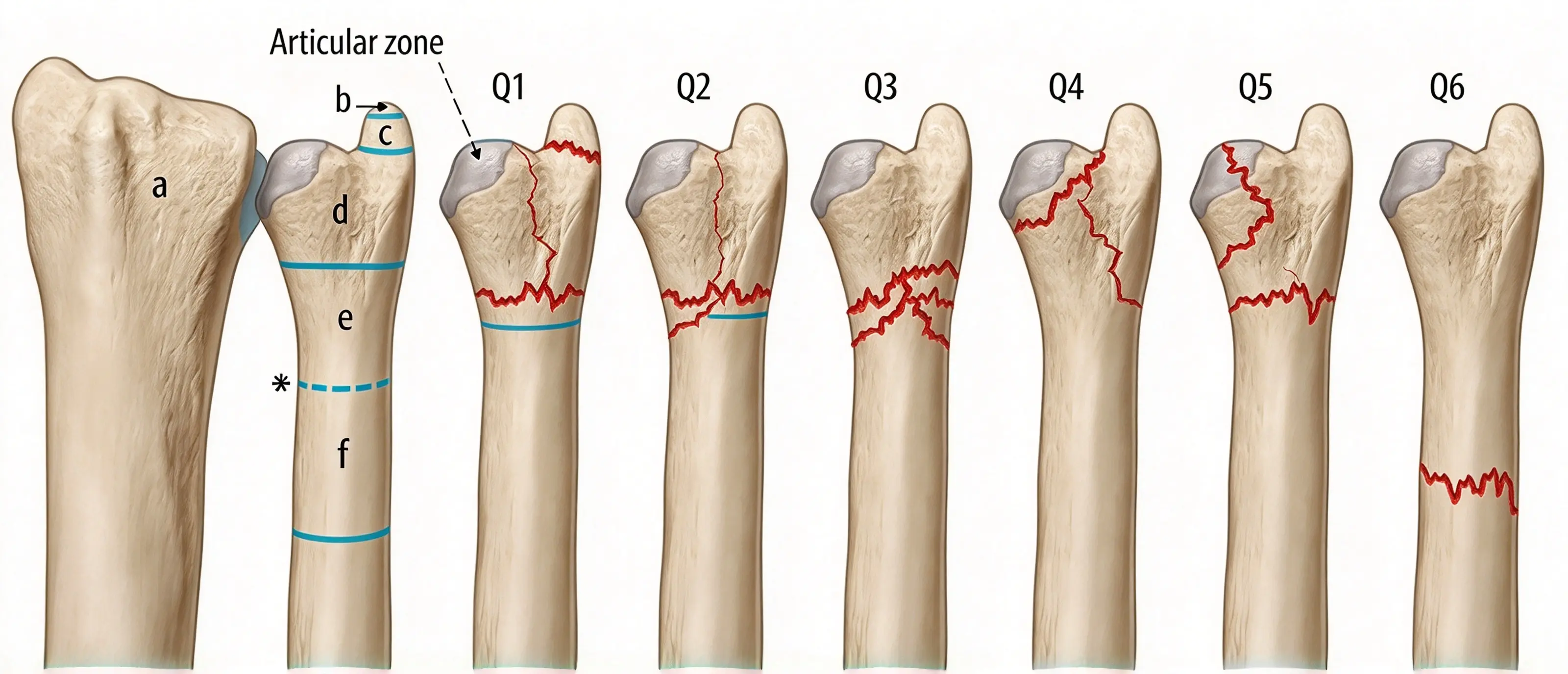
The source article describes six anatomical fracture patterns:
Q1: Ulnar styloid fracture
Q2: Simple ulnar neck fracture
Q3: Comminuted ulnar neck fracture
Q4: Ulnar head fracture
Q5: Combined ulnar head and neck fracture
Q6: Distal ulnar shaft fracture
These labels should be retained when reproducing the original classification illustration. They should not be presented as a universally accepted classification system unless the original publication establishing these categories is identified.
Fracture location alone does not determine the need for fixation. Displacement, articular involvement, fracture stability, DRUJ congruity, associated distal radius injury, bone quality, and functional demands must also be assessed.
The optimal management of distal ulna fractures remains debated. In patients with an associated distal radius fracture, restoration of radial alignment may improve DRUJ stability through the surrounding soft tissues, including the distal oblique bundle of the interosseous membrane.
Consequently, the need for direct ulnar fixation should be assessed after the distal radius has been reduced and stabilized when indicated.
If the DRUJ remains stable and the distal ulna fracture is acceptably aligned, additional fixation may not be necessary. Conversely, persistent instability, articular incongruity, or substantial displacement may justify surgical treatment.
The following features have been described as potential indications for distal ulna fracture fixation:
Angular deformity of 10° or more
Ulnar shortening of 3 mm or more
Fracture displacement exceeding one-third of the shaft diameter
Instability of the ulnar head or neck
Movement of fracture fragments during passive forearm rotation
Articular displacement or incongruity
Persistent DRUJ instability after treatment of an associated distal radius fracture
These numerical thresholds are reference criteria reported in the source material, not universal surgical rules. The decision to operate should consider the specific fracture pattern, joint stability, patient characteristics, and available clinical evidence.
Distal ulna ORIF may be considered in the following situations:
Displaced or unstable fractures
Fractures involving the ulnar head or neck may require fixation when instability or fragment displacement compromises alignment or joint function, whether or not a distal radius fracture is present.
DRUJ articular incongruity
Displacement involving the articular surface may interfere with DRUJ congruity and forearm rotation.
Open fractures
Open fractures may require debridement, fracture stabilization, and appropriate soft-tissue management. Definitive internal fixation depends on contamination, tissue viability, fracture stability, and the overall treatment plan. An open fracture does not automatically require immediate plating in every circumstance.
Bilateral injuries or multiple trauma
When both upper limbs are injured, or when associated injuries make prolonged immobilization especially problematic, stable fixation may help facilitate rehabilitation. The benefit must be balanced against the patient's overall medical condition.
High functional demands
Patients with substantial occupational, sporting, or daily functional requirements may benefit from restoration of alignment and stability when the fracture pattern and clinical assessment support fixation.
The source article identifies several situations in which distal ulna plating may be inappropriate or require additional consideration:
Associated injuries of the same limb that prevent early active rehabilitation, reducing the immediate functional benefit of fixation
Stable, nondisplaced fractures that do not require surgical stabilization
Severe osteoporosis of the ulnar head that prevents reliable screw purchase
Situations requiring a wrist-spanning plate or external fixation construct for the broader injury
These factors should not all be interpreted as absolute contraindications. For example, severe osteoporosis may increase fixation difficulty but does not automatically exclude surgery. Treatment should be individualized according to fracture morphology, available fixation options, soft-tissue conditions, and the patient's goals.
The ulnar approach provides access to the distal ulna while allowing the surgeon to identify and protect important neurovascular and tendinous structures.
The patient is positioned and prepared according to the planned procedure and associated injuries.
Before incision, the surgeon palpates the ulnar styloid to establish the distal reference point and the ulnar border or crest approximately 5 cm proximally to establish the proximal reference point.
A longitudinal incision approximately 5 cm long is made between the extensor carpi ulnaris (ECU) and flexor carpi ulnaris (FCU), beginning at the level of the ulnar styloid.
The exact incision length and position should be adjusted according to the fracture location, required exposure, and implant design.
Protection of the dorsal branch of the ulnar nerve (DBUN) is one of the most important safety considerations during distal ulna exposure.
The DBUN supplies sensation to part of the dorsoulnar hand. Injury can result in sensory disturbance, painful neuroma formation, or persistent neuropathic symptoms.
The nerve may emerge from the dorsal border of the FCU approximately 5 cm proximal to the pisiform, then travel through the subcutaneous tissues toward the dorsoulnar hand. Its course varies between individuals.
To reduce the risk of injury:
Limit the depth of the initial incision, particularly near its distal end.
Use skin retractors to improve visualization.
Carefully separate the subcutaneous tissues with blunt dissection until the fascia is identified.
Identify the DBUN before proceeding with deeper exposure.
Avoid excessive traction or blind dissection around the nerve.
The nerve should be identified based on its actual intraoperative course rather than relying exclusively on an average anatomical distance.
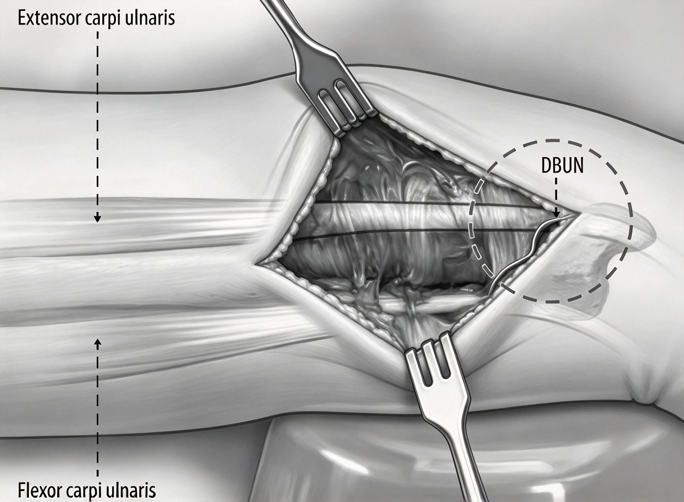
Once the relevant soft tissues have been protected, the fracture site is exposed sufficiently to permit reduction and fixation.
The exposure should preserve soft-tissue attachments where possible. Excessive dissection may compromise local blood supply and increase the risk of postoperative stiffness, tendon irritation, or wound complications.
The degree of exposure depends on whether the fracture involves the ulnar styloid, neck, head, or distal shaft and whether direct visualization of the articular surface is required.
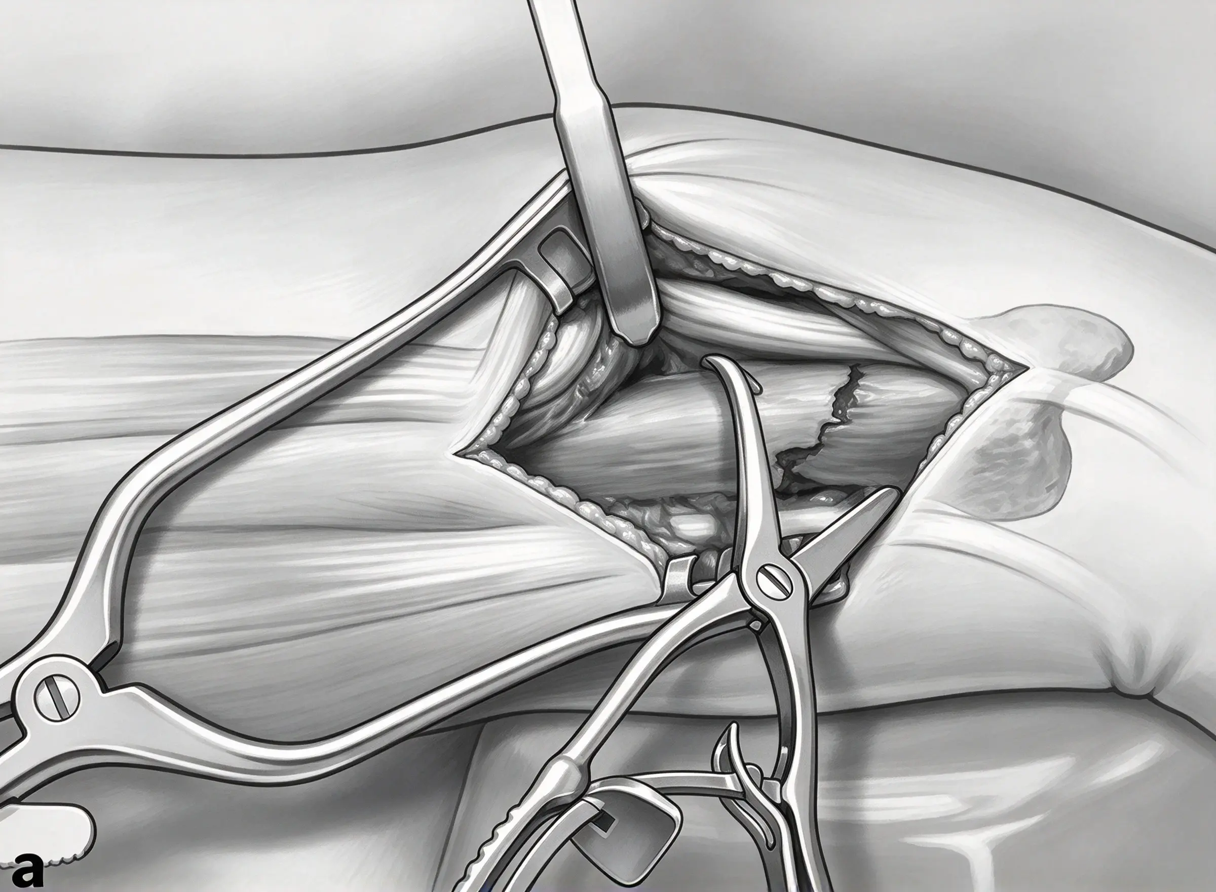
After exposure, the fracture is reduced using an appropriate reduction instrument, such as a Weber reduction clamp, according to the fracture configuration.
The objectives are to restore acceptable alignment, correct shortening and angular deformity, and achieve congruity of the articular surfaces where involved.
Depending on the fracture pattern, temporary K-wire fixation may help maintain the distal fragment in the desired position.
In selected cases, temporary fixation directed toward the distal radius may assist reduction or keep the fragment stable while the definitive plate is positioned. This can be particularly helpful when a reduction clamp interferes with plate placement.
Temporary fixation must be planned carefully to avoid injury to articular surfaces, tendons, nerves, or other structures. Fluoroscopic confirmation should be obtained before proceeding.
Intraoperative fluoroscopy is used to assess fracture alignment and articular congruity.
The surgeon should also evaluate DRUJ congruity, especially when the fracture is associated with a distal radius injury.
The reduction should be reassessed after temporary fixation and again after definitive plate and screw placement, because manipulation during fixation can cause loss of alignment.
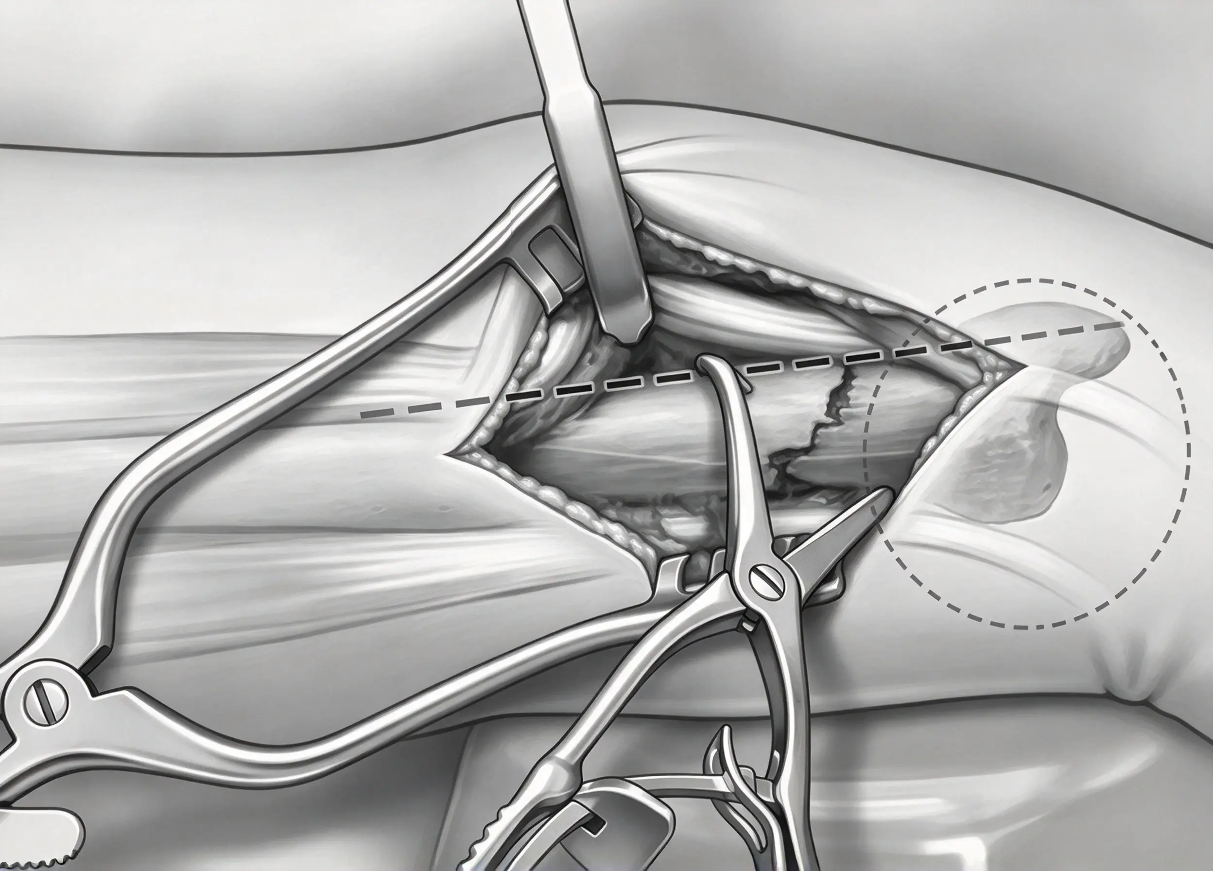
Correct plate positioning is critical to minimize irritation of the ECU tendon, avoid impingement, preserve forearm rotation, and achieve stable fixation.
The source article describes a relatively safe plate-placement region along the ulnar border and a small adjacent volar portion of the distal ulna.
Using the ulnar border aligned with the ulnar styloid as the 0° reference, the described safe zone extends approximately from 0° to 30° toward the volar side.
This anatomical reference is intended to help guide plate placement, but the actual safe region depends on individual anatomy, fracture morphology, plate geometry, and the position of surrounding tendons.
The source describes using a 1.5 mm or 2.0 mm locking compression plate (LCP), selected according to the fracture and available implant system.
The appropriate plate should provide sufficient fixation while minimizing prominence and interference with surrounding structures.
For fractures requiring very distal plate placement, a plate with a maximum width of approximately 20 mm, positioned on the volar aspect where anatomically suitable, may help reduce impingement. This dimension is specific to the described technique and should be verified against the actual implant design.
Volar placement may be preferable when it allows adequate fixation without interference with the ECU tendon.
If dorsal placement is necessary, the plate may be positioned immediately adjacent to the volar side of the ECU tendon, according to the described technique. Particular care is required around the ECU subsheath.
Patients should be informed when the selected plate position may increase the risk of tendon irritation, instability, or other complications.
Plate position should never be determined by the fracture line alone. The surgeon must balance access to the fracture, screw trajectories, fixation stability, tendon clearance, and preservation of forearm rotation.
The following sequence summarizes the technique described in the source article. It is intended for professional education and must be adapted to the selected implant system and the patient's anatomy.
For comminuted fractures, plate contouring may be necessary to achieve an appropriate fit against the bone.
The objective is to maintain a low-profile construct while obtaining adequate fixation. Excessive contouring or repeated bending may compromise implant integrity and should follow the manufacturer's instructions.
The described technique uses a 1.8 mm drill to prepare the initial hole, followed by a 2.4 mm cortical screw to secure the proximal portion of the plate.
This initial fixation point can allow the plate to rotate around the screw, facilitating fine adjustment of the distal plate position.
Drill diameter, screw diameter, and drilling technique must be verified for the specific plate system.
After the initial proximal screw has been inserted, the plate position can be adjusted to optimize distal coverage, screw trajectories, and clearance from the ECU tendon.
The surgeon should confirm that the plate remains within the intended safe region and does not interfere with surrounding soft tissues.
Once the plate position is satisfactory, variable-angle (VA) locking screws are inserted into the distal fragment according to the implant design.
Variable-angle screws can provide flexibility in screw trajectory, which may be useful when the distal fragment is small or comminuted. However, the selected trajectories must provide secure fixation without entering the DRUJ or other articular surfaces.
Fluoroscopy is used to evaluate fracture alignment, ulnar height, and the relationship between the distal radius and ulna.
If the reduction remains uncertain, the source article recommends comparing the injured side with preoperative radiographs of the uninjured side obtained in comparable forearm rotation positions.
Contralateral comparison can provide useful anatomical context, but normal variation and differences in positioning must be considered.
After satisfactory plate positioning has been confirmed, definitive fixation is completed.
The described construct uses:
Two proximal cortical screws
One proximal variable-angle locking screw
Four distal variable-angle locking screws
This is the configuration reported in the source technique, not a universal screw count. The actual number, size, and trajectory of screws must be selected according to fracture morphology, available bone stock, plate design, and manufacturer instructions.
Screw penetration into the ulnar head and DRUJ must be avoided.
Fluoroscopy should be used to assess screw length and trajectory. Multiple views may be needed because a screw that appears extra-articular on one projection may penetrate the joint on another.
The surgeon should confirm that the final construct provides adequate fixation without articular penetration or interference with adjacent structures.
After fixation, the wrist and forearm are moved through the available range of motion under direct observation and, where appropriate, fluoroscopic guidance.
The assessment should identify:
Restriction of pronation or supination
Plate or screw impingement
ECU tendon irritation or instability
Abnormal contact between the plate and surrounding tissues
Loss of reduction during movement
If motion is restricted or impingement is suspected, plate position and screw trajectories should be reassessed before closure.
The source technique recommends obtaining intraoperative images in pronation, neutral rotation, and supination.
These views can help identify screw penetration, altered joint congruity, and position-dependent impingement that may not be apparent in a single image.
Before wound closure, DRUJ stability should be evaluated using a controlled manual push-pull or ballottement test.
The assessment should consider the endpoint of translation and compare the injured side with the contralateral wrist when appropriate. Comparison is particularly useful because normal DRUJ laxity varies between individuals.
If instability persists after fracture fixation, additional treatment may be required depending on the injury pattern. Options described in the source include fixation of a relevant ulnar styloid fracture or temporary transfixion of the distal ulna to the radius with K-wires in a stable rotational position.
The appropriate method depends on the underlying cause of instability, the condition of the TFCC, fracture morphology, and the stability achieved after reduction.
If the DRUJ is stable and the fixation construct is satisfactory, the wound can be irrigated and closed using an appropriate closure technique.
Several technical considerations are particularly important when performing distal ulna ORIF.
Radiographs should be obtained before surgery to characterize the fracture and assist planning. Intraoperative imaging is necessary to confirm reduction and implant position. Postoperative imaging helps document the final construct and provides a baseline for follow-up.
The imaging protocol should be appropriate to the fracture pattern and clinical circumstances.
Avoid excessive traction on the distal soft tissues during exposure. The DBUN may be superficial and vulnerable to injury, especially near the distal end of the incision.
Direct identification and careful retraction are preferable to blind dissection.
The plate should be positioned to provide adequate fixation without restricting forearm rotation or irritating the ECU tendon.
The surgeon should inspect tendon clearance and assess movement before closure. If the plate interferes with the tendon or restricts rotation, its position should be reconsidered.
Comminuted fractures can make restoration of ulnar length difficult. When the anatomy is uncertain, comparison with the uninjured side may help assess ulnar variance and alignment.
This is especially important when an associated distal radius fracture has altered radial height, because restoring the distal ulna without accounting for radial shortening may produce relative positive ulnar variance and increase the risk of ulnocarpal impaction.
The DRUJ should be assessed in pronation, neutral rotation, and supination as appropriate.
A controlled manual push-pull test, combined with assessment of joint congruity and the quality of the endpoint, can help identify residual instability.
Comparison with the opposite wrist can improve interpretation, although the clinical findings must be considered alongside imaging and the overall injury pattern.
Complications may result from the initial injury, fracture reduction, implant positioning, or postoperative healing. Understanding the mechanisms of failure helps guide prevention.
Injury to the DBUN may cause numbness, altered sensation, neuropathic pain, or a painful neuroma along the dorsoulnar hand.
Risk reduction: Identify the nerve during exposure, minimize blind dissection, and avoid excessive traction or compression during retraction and closure.
A screw that penetrates the ulnar head into the DRUJ can damage the articular surface and restrict forearm rotation.
Risk reduction: Confirm screw length and trajectory using appropriate fluoroscopic views. Assess the construct in multiple projections rather than relying on a single image.
Residual displacement or articular incongruity can compromise DRUJ mechanics and limit pronation or supination.
Risk reduction: Evaluate reduction before definitive fixation and reassess alignment after the plate and screws are inserted. Use additional imaging or contralateral comparison when necessary.
Overcorrection or undercorrection of ulnar length may alter load distribution and contribute to ulnocarpal impaction or DRUJ-related symptoms.
An associated distal radius fracture adds complexity because loss of radial height can change the relative relationship between the radius and ulna.
Risk reduction: Assess both bones and their relationship. Confirm ulnar height and variance after reduction rather than evaluating the ulna in isolation.
A prominent plate or unsuitable plate position can cause pain, tendon irritation, or restriction of forearm rotation.
Risk reduction: Select an appropriate low-profile plate, assess the anatomical safe zone, and test wrist and forearm motion before closure.
Damage to the ECU subsheath may lead to tendon instability or subluxation during forearm rotation and wrist movement.
Risk reduction: Identify the ECU tendon and its surrounding structures during exposure. Avoid unnecessary disruption of the subsheath and assess tendon stability after fixation.
DRUJ instability may persist despite distal ulna fixation, particularly when the injury also involves the TFCC or other stabilizing structures.
Risk reduction: Perform a controlled stability assessment after fixation. If instability remains, investigate the underlying cause and consider additional treatment based on the injury pattern.
Inadequate fixation, poor bone quality, residual instability, or loss of reduction can lead to secondary displacement or implant failure.
Risk reduction: Obtain appropriate reduction, select a fixation construct suitable for the available bone stock, verify screw placement, and follow the postoperative imaging and rehabilitation plan.
Postoperative care should protect the fixation construct while restoring wrist and forearm function.
The rehabilitation plan depends on fracture stability, fixation quality, associated distal radius injury, soft-tissue condition, and any additional procedures performed to stabilize the DRUJ.
Key considerations include:
Monitoring the wound and neurovascular status
Assessing pain, swelling, and finger mobility
Following the surgeon's instructions for immobilization and activity
Initiating wrist and forearm motion when permitted by the fixation construct and associated injuries
Avoiding premature loading or movement that could jeopardize an unstable repair
Arranging follow-up imaging to assess alignment and healing
Monitoring for tendon irritation, nerve symptoms, joint stiffness, or persistent instability
Early active movement may be a goal of stable fixation, but it should not be assumed to be safe in every case. Rehabilitation timing must be individualized.
No. Stable, nondisplaced fractures may be treated without direct fixation. Surgery is more likely to be considered when the fracture is displaced, unstable, involves the articular surface, or contributes to persistent DRUJ instability.
Not necessarily. Fixation of the distal radius may restore DRUJ stability in some patients. The need for direct ulnar fixation should be reassessed after the radius has been reduced and stabilized, taking fracture alignment and joint stability into account.
The plate should be positioned according to the fracture pattern, implant design, and surrounding anatomy. The described technique identifies the ulnar border and a limited adjacent volar region as a potential safe zone. The surgeon must confirm adequate fixation while avoiding ECU tendon irritation, impingement, and articular screw penetration.
The DBUN is superficial near the operative field and may be injured during incision, dissection, or retraction. Injury can cause sensory disturbance or a painful neuroma. Careful identification and protection are therefore essential.
A controlled manual push-pull or ballottement test is used to assess translation and the quality of the endpoint. Assessment in different forearm rotational positions and comparison with the contralateral wrist can help identify residual instability.
Important complications include DBUN injury, intra-articular screw penetration, inadequate reduction, abnormal ulnar variance, plate impingement, ECU tendon irritation or instability, persistent DRUJ instability, and fixation failure.
Distal ulna fracture fixation requires careful evaluation of fracture morphology, DRUJ stability, associated distal radius injuries, and the patient's functional needs. Although not every distal ulna fracture requires surgery, displaced or unstable fractures and those associated with persistent joint incongruity may benefit from ORIF.
A successful procedure depends on safe surgical exposure, protection of the dorsal branch of the ulnar nerve, accurate reduction, appropriate plate positioning, secure screw fixation, and confirmation that the construct does not interfere with the ECU tendon or DRUJ.
Intraoperative imaging and assessment of forearm rotation are essential for identifying technical problems before closure. A final stability examination helps determine whether additional treatment is needed for persistent DRUJ instability.
By combining appropriate patient selection, careful surgical technique, and individualized postoperative rehabilitation, clinicians can work toward restoring alignment, maintaining joint stability, and improving functional recovery after distal ulna fractures.
Medical Disclaimer: This article is intended for educational purposes and does not replace professional clinical judgment, individualized surgical planning, or the instructions for use provided with a specific implant system. Surgical indications, implant selection, screw configuration, and rehabilitation protocols must be determined by qualified healthcare professionals.


