
Views: 0 Author: Site Editor Publish Time: 2026-09-28 Origin: Site
Introduction
Accurate identification of ligament attachment sites is fundamental to anatomic knee ligament reconstruction. The position of a femoral or tibial tunnel can substantially influence graft orientation, tension, joint kinematics, and postoperative stability. For this reason, surgeons performing ligament reconstruction increasingly rely on a combination of three-dimensional anatomy, preoperative imaging, intraoperative bony landmarks, and quantitative measurements.
A JBJS review by Asheesh Bedi and colleagues systematically examined the anatomy and imaging landmarks of six major ligamentous structures around the knee:
Anterior cruciate ligament (ACL)
Posterior cruciate ligament (PCL)
Medial collateral ligament (MCL)
Lateral collateral ligament (LCL)
Anterolateral ligament (ALL)
Medial patellofemoral ligament (MPFL)
The review summarized the major osseous landmarks, ligament footprints, and radiographic measurements that can help surgeons identify native attachment sites.
Importantly, these measurements should be regarded as anatomic reference values rather than rigid surgical coordinates. Individual variation in bone size, morphology, ligament footprint, previous surgery, and injury pattern must be considered during reconstruction.
This article reviews the key anatomy and imaging landmarks of the six structures and discusses how surgeons can integrate imaging-based planning with intraoperative anatomic identification.
The major ligaments of the knee work together to control anterior-posterior translation, varus-valgus stability, and rotational motion.
When a ligament is reconstructed, the objective is generally to reproduce the native attachment and functional orientation as closely as appropriate for the individual patient.
An incorrectly positioned tunnel may alter:
Graft length and tension
Graft orientation
Joint kinematics
Rotational stability
Contact mechanics
Graft loading
Risk of residual instability
Risk of reconstruction failure
For this reason, anatomic reconstruction is not simply a matter of drilling a tunnel at a fixed percentage of the bone.
Quantitative radiographic methods are valuable because they provide reproducible reference points. However, these measurements should be combined with direct identification of native bony landmarks and ligament footprints during surgery.
A practical workflow is therefore:
Preoperative imaging → quantitative localization → intraoperative anatomic verification → tunnel preparation → postoperative imaging assessment
The ACL originates from the medial aspect of the lateral femoral condyle and extends to the anterior intercondylar region of the tibia.
It is commonly described as having two major functional bundles:
Anteromedial (AM) bundle
Posterolateral (PL) bundle
The names reflect their relative positions on the tibial footprint.
The ACL contributes substantially to restraint of anterior tibial translation and also participates in control of tibial rotation.
The ligament has an average length of approximately 31 ± 2 mm and a mid-substance diameter of approximately 10 ± 2 mm in commonly cited anatomic studies. Its footprint expands substantially toward the bone, producing a broad attachment rather than a simple cylindrical insertion.
Two important bony landmarks can be identified on the medial wall of the lateral femoral condyle:
The lateral intercondylar ridge, often called the resident's ridge, forms an important proximal/anterior boundary of the ACL femoral footprint.
It generally runs from posteroproximal to anterodistal across the femoral attachment area.
The lateral bifurcate ridge runs approximately perpendicular to the intercondylar ridge and separates the AM and PL bundle attachment regions.
These landmarks can therefore help identify the native ACL footprint during anatomic reconstruction.
Reported measurements place the center of the AM bundle approximately 4.8 mm proximal to the bifurcate ridge and 7.1 mm posterior to the intercondylar ridge, while the PL bundle center is approximately 5.2 mm distal to the bifurcate ridge and 3.6 mm posterior to the intercondylar ridge.
The overall ACL footprint center has also been reported approximately 1.7 mm proximal to the bifurcate ridge and 6.1 mm posterior to the intercondylar ridge.
Because these values vary between studies and specimens, they should be treated as reference measurements rather than universal drilling coordinates.
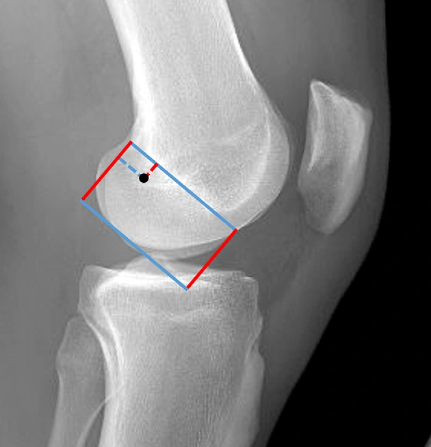
Figure 1. Bernard-Hertel quadrant localization method for the femoral attachment of the anterior cruciate ligament
The Bernard-Hertel quadrant method provides a radiographic method for describing the femoral ACL footprint.
The medial wall of the lateral femoral condyle can be divided using:
The Blumensaat line
A line perpendicular to the Blumensaat line
The anterior-posterior dimension of the condyle
The proximal-distal dimension of the condyle
The ACL footprint center has been described near the intersection of the first and second quadrants.
Reported measurements place the footprint center approximately:
28.5% of the Blumensaat line depth from the posterior reference
24.8% of the condylar height from the distal reference
For double-bundle reconstruction, the AM and PL bundle locations can also be expressed as percentages along the sagittal dimension of the femoral footprint.
These percentage-based methods are particularly useful when reviewing preoperative and postoperative lateral radiographs.
The tibial ACL footprint occupies the anterior intercondylar region between the medial and lateral tibial plateaus.
Several important landmarks can assist with localization.
The center of the ACL tibial footprint has been described approximately:
7.5 mm medial to the anterior horn of the lateral meniscus
13.0 mm anterior to the posterior intercondylar ridge
10.5 mm anterior to the ACL ridge
The posterior margin of the ACL footprint is related to the intercondylar eminence and the ridge between the medial and lateral intercondylar tubercles.
An important anatomical feature is the overlap between the ACL footprint and the anterior horn of the lateral meniscus.
Studies have reported substantial overlap in both coronal and sagittal dimensions.
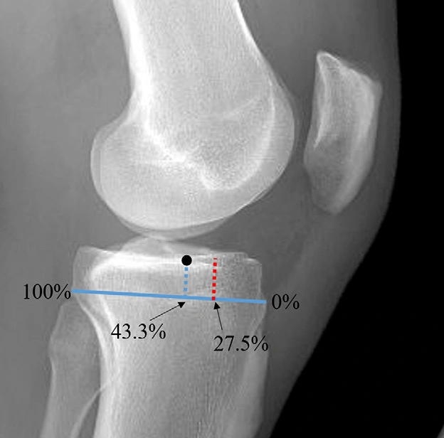
Figure 2. Anteroposterior percentage localization method for the tibial attachment of the anterior cruciate ligament
The ACL tibial footprint center has been reported at approximately 43.3% of the anteroposterior dimension of the tibial plateau, using the posterior plateau as the 100% reference.
The most anterior ACL fibers may extend to approximately 27.5% of the tibial plateau depth, although the exact footprint varies between individuals.
In double-bundle descriptions, the AM and PL bundles can also be localized using mediolateral and anteroposterior percentage measurements.
These values can help with preoperative planning, but direct visualization of the native footprint remains important whenever feasible.
The PCL extends from the posterior tibial intercondylar region to the medial femoral condyle.
It is the primary restraint to posterior tibial translation and also contributes to rotational stability.
Two major functional components are commonly described:
Anterolateral (AL) bundle
Posteromedial (PM) bundle
The PCL is not isometric throughout the range of motion.
The AL bundle tends to become more taut with knee flexion, whereas the PM bundle contributes more strongly near knee extension.
The PCL is approximately 38 mm long in commonly cited anatomic studies, with a mid-substance diameter of approximately 13 mm.
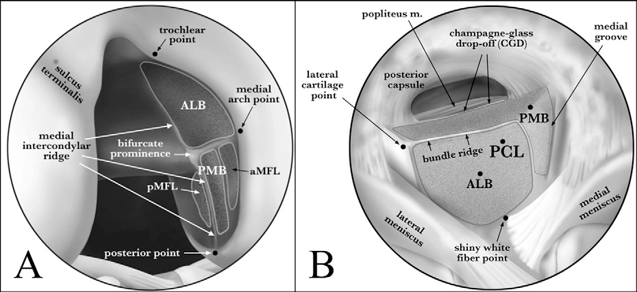
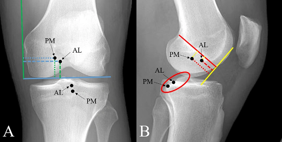
Figure 4. Anteroposterior and lateral imaging localization of the posterior cruciate ligament attachments
Note: 4-A Anteroposterior radiograph. The green solid line indicates the medial epicondyle line, and the blue solid line indicates the vertical line to the distal femoral condyle, marking the distance parameters of the anterolateral (AL) and posteromedial (PM) bundles; 4-B Lateral radiograph. The red solid line indicates the Blumensaat line, and the yellow solid line indicates its perpendicular line. The red circle indicates the PCL articular surface, and the center of the tibial attachment of the ligament is located 7 mm proximal between the two bundles on this surface.
Similar to the ACL, important bony ridges help identify the PCL femoral footprint.
The medial intercondylar ridge and bifurcate ridge are useful landmarks.
The bifurcate ridge separates the attachment regions of the two major PCL bundles.
The AL and PM bundle centers have been reported to be approximately 12.1 ± 1.3 mm apart.
The attachment regions are located at different distances from the articular cartilage margin, providing useful references during anatomic PCL reconstruction.
On anteroposterior imaging, the AL and PM bundle centers can be described relative to the medial femoral condyle.
Reported measurements include approximately:
34.1 ± 3.0 mm from the most medial aspect of the medial femoral condyle to the AL bundle center
29.2 ± 3.0 mm to the PM bundle center
Their relationship to the distal femoral reference can also be quantified.
On lateral imaging, measurements can be made relative to the Blumensaat line and a perpendicular reference line extending from the anterior femoral cortex.
These measurements provide a framework for evaluating the femoral position of PCL reconstruction tunnels.
The tibial PCL footprint occupies the posterior intercondylar region.
Important arthroscopic landmarks include:
Lateral tibial plateau cartilage
Posterior root of the medial meniscus
Interbundle ridge
Posterior tibial cortex
Popliteus region
The PCL tibial attachment is closely related to the posterior tibial plateau and extends toward the posterior tibial surface.
The center of the tibial footprint has been described approximately 7 mm from the posterior tibial cortex in certain anatomic studies.
The AL and PM components have different relationships to the tibial articular surface.
During PCL reconstruction, understanding these relationships is particularly important because the tibial tunnel passes through a region where inaccurate positioning may affect graft trajectory and increase the risk of tunnel-related complications.
The MCL, also known as the tibial collateral ligament, is the primary static restraint to valgus stress at the knee.
It is commonly divided into:
Superficial MCL
Deep MCL
The superficial MCL is the larger and more structurally independent component and is frequently the principal structure considered during MCL reconstruction.
The deep MCL is closely associated with the joint capsule and medial meniscus.
Unlike the cord-like LCL, the superficial MCL has a broad, relatively flat morphology.
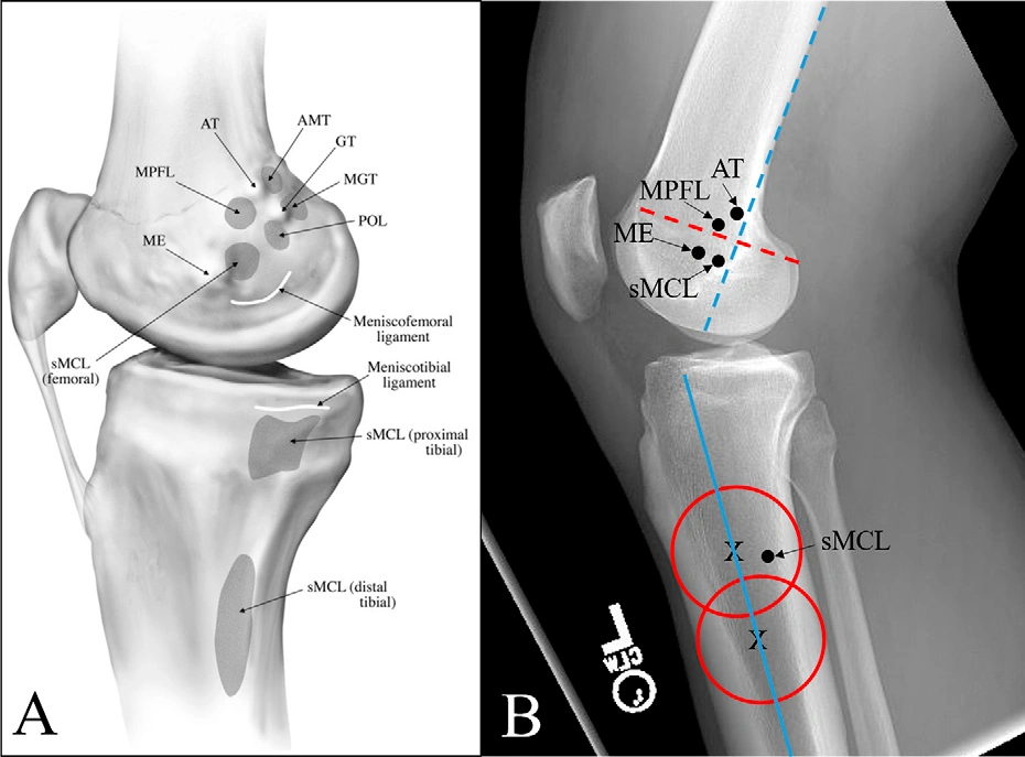
Figure 5. Anatomy and imaging localization of the medial structures and ligament attachments of the knee
Note: 5-A shows the medial anatomical structures of the knee. AT indicates the adductor tubercle, ME indicates the medial epicondyle, sMCL indicates the superficial medial collateral ligament, and MPFL indicates the medial patellofemoral ligament; 5-B shows the imaging localization lines. The blue dashed line indicates the posterior femoral cortex line, the red dashed line indicates its perpendicular line, and the blue solid line indicates the tibial shaft axis, marking the relative positions of the structures.
The superficial MCL femoral attachment is located near the medial epicondyle.
A commonly cited location is approximately:
4.8 mm posterior to the medial epicondyle
3.2 mm proximal to the medial epicondyle
The direct distance between the epicondyle and the MCL attachment has been reported at approximately 6 mm in anatomic studies.
Radiographically, the attachment can also be localized relative to the posterior femoral cortex and the Blumensaat line.
The tibial MCL attachment is substantially broader than its femoral attachment.
The main distal attachment has been reported approximately 61 mm distal to the joint line, with another more proximal attachment region approximately 16 mm distal to the joint line.
The distal superficial MCL attachment extends along the posteromedial tibia and contributes to the formation of the floor of the pes anserine region.
Because the tibial footprint is broad, reconstructive procedures should account for the native attachment rather than treating the MCL as a simple point-to-point structure.
The LCL, also known as the fibular collateral ligament, is the primary static restraint to varus stress at the knee.
Unlike the broad MCL, the LCL is a relatively narrow, cord-like structure.
It is approximately 3–4 mm in diameter and approximately 66 mm long in commonly reported anatomic measurements.
The LCL also contributes to rotational stability.
Its length changes relatively little through much of the early range of knee flexion, with greater shortening occurring at higher flexion angles.
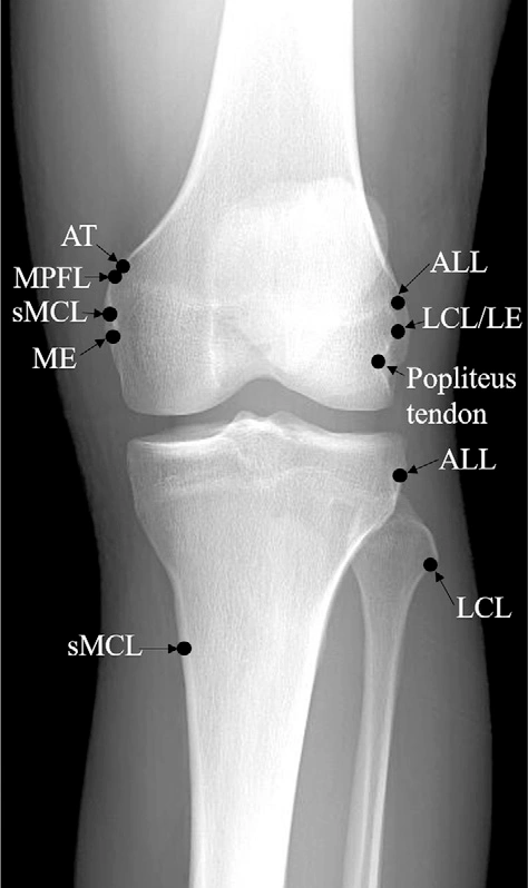
Figure 6. Imaging attachment localization of the medial and lateral periarticular structures of the knee
Note: AT indicates the adductor tubercle, MPFL indicates the medial patellofemoral ligament, sMCL indicates the superficial medial collateral ligament, ME indicates the medial epicondyle, ALL indicates the anterolateral ligament, LCL indicates the lateral collateral ligament, and LE indicates the lateral epicondyle, marking the relative positions of the ligaments on the anteroposterior radiograph and their distances from the joint line.
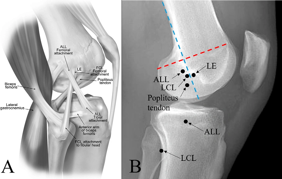
Figure 7. Anatomy and imaging localization of the lateral structures and ligament attachments of the knee
Note: 7-A shows the lateral anatomical structures of the knee. LE indicates the lateral epicondyle, ALL indicates the anterolateral ligament, and LCL indicates the lateral collateral ligament; 7-B shows the imaging localization of the lateral ligaments, marking the relative positions of the attachments of the anterolateral ligament and the lateral collateral ligament.
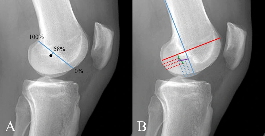
Figure 8. Two imaging localization methods for the femoral attachment of the lateral collateral ligament
Note: 8-A is the Blumensaat line percentage method. The black dot indicates the center of the attachment, located at 58% ± 4.7% of the lateral femoral condylar width along this line and 2.3 ± 2.3 mm distal to it; 8-B is the Meister method. The blue solid line indicates the posterior border of the femoral shaft, and the red solid line indicates its perpendicular line. A point at 27% of its length is marked, and the intersection of the curves is the center of the attachment.
The femoral attachment of the LCL is located near the lateral epicondyle.
A commonly described attachment site is approximately:
3.1 mm posterior to the lateral epicondyle
1.4 mm proximal to the lateral epicondyle
The attachment is associated with a small bony depression near the lateral femoral epicondyle.
It is also located in relation to the popliteus tendon attachment, providing an additional landmark during lateral-sided knee reconstruction.
On anteroposterior radiographs, the femoral attachment can be described relative to the distal femoral reference.
On lateral radiographs, the LCL attachment has been described at approximately 58% of the mediolateral width of the lateral femoral condyle, with additional measurements relative to the Blumensaat line.
Another method uses the posterior femoral cortical line and a perpendicular reference to identify the attachment.
These radiographic techniques can help verify the location of a femoral tunnel during postoperative assessment.
The distal LCL attachment is located on the fibular head.
The attachment occupies a characteristic area on the anterosuperior portion of the fibular head.
An important surgical landmark is the bony platform on the fibular head where the LCL attaches.
Identification of this structure is particularly important during lateral collateral ligament and posterolateral corner reconstruction because several important structures converge around the fibular head.
The common peroneal nerve also courses around the fibular neck and must be carefully protected during lateral knee surgery.
The anterolateral ligament remains one of the more debated structures in contemporary knee anatomy.
Some anatomical studies have identified it as a distinct ligamentous structure, whereas other studies have described it as part of the anterolateral capsule or a capsular thickening.
Reported measurements vary considerably depending on how the structure is defined.
This variation is important when interpreting published measurements or planning an anterolateral reconstruction.
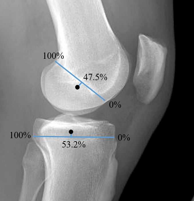
Figure 9. Lateral radiographic localization of the anterolateral ligament
Note: The upper image shows the femoral attachment, located at 47.5% ± 4.3% posterior to the anterior border of the femoral condyle and 3.7 ± 1.1 mm below the Blumensaat line; the lower image shows the tibial attachment, located at 53.2% ± 5.8% posterior to the anterior border of the lateral tibial plateau, with 100% representing the anteroposterior diameter distance of the corresponding border.
The femoral attachment of the ALL has been described near the lateral epicondylar region, posterior and proximal to the LCL femoral attachment.
One commonly cited anatomical relationship places the ALL femoral attachment approximately 4.7 mm posterior and 8.4 mm proximal to the lateral epicondyle.
On radiographs, its location can be expressed relative to:
The femoral joint line
The anterior border of the lateral femoral condyle
The Blumensaat line
Reported values place the femoral attachment approximately 15.8–22.3 mm proximal to the joint line on certain AP measurements.
The tibial attachment of the ALL is generally described on the anterolateral tibia between:
The Gerdy's tubercle region
The fibular head
The attachment is located distal to the lateral joint line.
A commonly reported reference is approximately 9.5 mm distal to the joint line, although published measurements vary.
On lateral imaging, the attachment has been described at approximately 53% of the anteroposterior width of the lateral tibial plateau, depending on the reference method.
The ALL has been investigated primarily in relation to rotational stability of the knee, particularly control of internal tibial rotation.
Biomechanical studies have reported that the anterolateral structures contribute to rotational stability, especially at higher degrees of knee flexion.
However, the precise contribution of the ALL relative to the ACL, lateral meniscus, capsule, iliotibial band, and other anterolateral structures remains an area of ongoing research.
Therefore, the ALL should be understood as part of a broader anterolateral stabilizing complex, rather than considered in isolation.
The medial patellofemoral ligament is a major soft-tissue restraint against lateral patellar displacement, particularly during the early phase of knee flexion before the patella becomes more deeply engaged within the trochlear groove.
The MPFL is commonly considered the principal passive soft-tissue stabilizer against lateral patellar translation in this range.
The adult MPFL has been reported to measure approximately 53–62 mm in length, with a broad attachment and an average mid-substance width of approximately 19 mm, although substantial anatomical variation exists.
The femoral attachment is located in the region between the:
Adductor tubercle
Medial epicondyle
It forms a relatively broad attachment zone rather than a simple point.
One commonly reported anatomical description places the MPFL attachment approximately:
1.9 mm anterior to the adductor tubercle
3.8 mm distal to the adductor tubercle
The attachment is therefore located within the characteristic groove between the adductor tubercle and medial epicondyle.
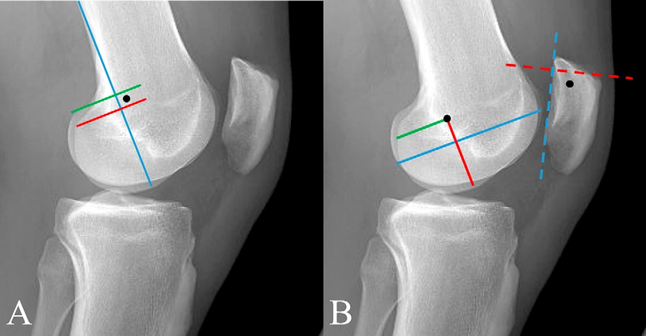
Figure 10. Imaging localization methods for the medial patellofemoral ligament
Note: 10-A is the Schöttle point localization. The blue line indicates the extension of the posterior femoral cortex, the red line indicates the perpendicular line at the most posterior point of the Blumensaat line, the green line indicates the perpendicular line at the proximal femoral condyle, and the black dot indicates the center of the attachment located 2 mm anterior to this region; 10-B is the percentage localization. The femoral attachment is located at 40% of the anteroposterior diameter and 50% of the proximodistal diameter of the medial femoral condyle, and the patellar attachment is marked in the proximal one-third region of the medial patella.
The Schöttle point is widely used as a radiographic reference for femoral MPFL reconstruction.
On a true lateral radiograph, the reference is based on the relationship among:
The posterior femoral cortical line
The Blumensaat line
A perpendicular reference from the proximal condyle
The commonly described target lies approximately 2 mm anterior to the intersection region defined by these reference lines.
The Schöttle point is useful because it provides a reproducible radiographic reference.
However, radiographic positioning and individual femoral morphology can influence the apparent location of the point. Therefore, a technically adequate lateral radiograph and careful anatomical assessment remain essential.
The patellar attachment is located primarily along the proximal third of the medial patella.
The attachment zone can extend approximately 17–30 mm, depending on the anatomical definition used.
An important anatomical variation is that MPFL fibers may extend into the medial quadriceps tendon.
Some anatomical studies have identified this quadriceps extension in a high proportion of specimens.
This finding is relevant because the native medial patellofemoral stabilizing structure is not always confined to a single isolated ligament band.
| Structure | Primary region | Major functional role | Important localization landmarks |
|---|---|---|---|
| ACL | Central knee | Controls anterior tibial translation and contributes to rotational stability | Resident's ridge, bifurcate ridge, tibial intercondylar region |
| PCL | Central/posterior knee | Primary restraint to posterior tibial translation | Medial intercondylar ridge, bifurcate ridge, posterior tibial footprint |
| MCL | Medial knee | Primary restraint to valgus stress | Medial epicondyle, posteromedial tibia |
| LCL | Lateral knee | Primary restraint to varus stress | Lateral epicondyle, fibular head |
| ALL | Anterolateral knee | Contributes to rotational stability | Lateral epicondylar region, Gerdy's tubercle/fibular head region |
| MPFL | Medial patellofemoral region | Major restraint to lateral patellar displacement | Adductor tubercle/medial epicondyle region, proximal medial patella |
The value of quantitative anatomical data is greatest when it is incorporated into a structured surgical workflow.
Evaluate:
Bone morphology
Existing tunnels
Previous hardware
Ligament injury pattern
Meniscal anatomy
Joint alignment
Cartilage condition
Associated fractures or bone loss
MRI can help identify the injured ligament and its native footprint, while CT may be particularly useful in revision cases for assessing tunnel position and bone morphology.
Use appropriate radiographic measurements to estimate the native attachment.
For example:
Bernard-Hertel quadrant method for ACL femoral localization
Percentage-based tibial measurements for ACL
Blumensaat-line relationships for cruciate ligament localization
Epicondylar landmarks for MCL and LCL
Schöttle point for MPFL reconstruction
Radiographic measurements should be confirmed against the patient's actual anatomy.
Useful intraoperative landmarks include:
Intercondylar ridges
Bifurcate ridges
Femoral epicondyles
Adductor tubercle
Tibial plateau
Meniscal roots
Gerdy's tubercle
Fibular head
A published measurement does not necessarily represent the exact footprint of every patient.
Factors affecting localization include:
Sex
Height
Bone size
Skeletal morphology
Ligament size
Previous injury
Previous reconstruction
Bone tunnel enlargement
Post-traumatic deformity
Therefore, quantitative measurements should support rather than replace anatomical judgment.
Anatomic landmarks and imaging measurements are particularly useful in revision surgery.
A revision reconstruction should begin with assessment of the existing tunnels.
CT and MRI can help determine:
Tunnel position
Tunnel widening
Tunnel convergence
Bone stock
Hardware position
Relationship between previous and planned tunnels
The anatomical reference values discussed above can then help determine whether an existing tunnel is close enough to the native footprint to be reused or whether a new tunnel or staged reconstruction may be necessary.
In multiligament reconstruction, tunnel planning becomes even more important because multiple tunnels may occupy relatively close anatomical regions.
When several ligaments are injured simultaneously, reconstruction must account for the three-dimensional relationship among the different attachment sites.
This is particularly relevant on the medial and lateral sides of the knee.
For example, the femoral attachment regions of the MCL and MPFL are anatomically close, meaning that tunnel position and trajectory must be planned carefully when both structures require reconstruction.
Likewise, reconstruction of the LCL and other posterolateral structures requires careful attention to the fibular head and lateral femoral anatomy.
A three-dimensional understanding of the attachment sites can reduce the risk of:
Tunnel convergence
Insufficient bone between tunnels
Graft malposition
Hardware conflict
Altered graft tension
A percentage-based measurement is a reference, not an absolute coordinate.
Radiographic magnification, knee rotation, bone morphology, and imaging quality can affect the apparent position.
Anatomic reconstruction requires recognition of the actual footprint and surrounding bony structures.
Radiographs should complement rather than replace intraoperative anatomy.
Several knee ligaments have broad or complex footprints.
This is particularly relevant for the ACL, MCL, and MPFL.
The same numerical measurement cannot be expected to apply identically to every patient.
In revision surgery, the existing tunnel may alter the available bone stock and may not correspond to the native footprint.
The ACL has a broad, anatomically defined femoral and tibial footprint. Tunnel position influences graft orientation and tension and can affect postoperative stability. Accurate localization is therefore an important component of anatomic ACL reconstruction.
The resident's ridge, or lateral intercondylar ridge, is a bony ridge on the medial wall of the lateral femoral condyle that helps define the ACL femoral footprint.
The bifurcate ridge is a bony landmark that separates the AM and PL regions of the ACL femoral footprint and can also be identified in relation to the PCL footprint on the medial femoral condyle.
It is a radiographic method used to describe the location of the ACL femoral footprint relative to the Blumensaat line and the dimensions of the lateral femoral condyle.
The Schöttle point is a radiographic reference used to estimate the femoral attachment of the MPFL during medial patellofemoral ligament reconstruction.
The anatomy of the ALL remains debated. Some studies describe it as a distinct ligament, while others consider it part of the anterolateral capsule or a capsular thickening. Its relationship with rotational stability is well studied, but its exact anatomical definition remains an area of research.
No. Measurements reported in anatomical and imaging studies should be considered reference values. Individual anatomy, imaging technique, skeletal size, previous injury, and reconstruction history can all influence the actual footprint.
Accurate localization of ligament attachment sites is a fundamental component of anatomic knee ligament reconstruction.
The ACL and PCL have identifiable intercondylar bony landmarks that can help surgeons distinguish their functional bundle regions. The MCL and LCL have characteristic femoral and tibial or fibular attachment sites around the epicondyles and proximal tibia or fibula. The ALL remains anatomically debated but is clinically relevant to the study of anterolateral rotational stability. The MPFL has a broad femoral attachment near the adductor tubercle and medial epicondyle and a proximal medial patellar attachment.
Quantitative imaging methods—including the Bernard-Hertel quadrant method, Blumensaat-line measurements, percentage-based tibial localization, epicondylar reference measurements, and the Schöttle point—can provide valuable assistance in preoperative planning and postoperative assessment.
However, these measurements should not be applied mechanically. The most reliable approach combines preoperative imaging, quantitative reference values, direct anatomical identification, individual patient morphology, and intraoperative assessment.
For surgeons performing primary, revision, or multiligament knee reconstruction, understanding the relationship between radiographic landmarks and native ligament footprints can help create a more systematic approach to tunnel planning and anatomical reconstruction.
Bedi A, et al. JBJS review of the anatomy and imaging landmarks of the major knee ligaments.
Bernard M, Hertel P. Radiographic localization of the ACL femoral insertion using the quadrant method.
Schöttle PB, et al. Radiographic localization of the femoral attachment for medial patellofemoral ligament reconstruction.
Anatomical and biomechanical studies of the ACL, PCL, MCL, LCL, ALL, and MPFL.
Contemporary literature on anatomic and multiligament knee reconstruction.


