Views: 0 Author: Site Editor Publish Time: 2026-09-10 Origin: Site
Metacarpal Fracture Treatment: A Comprehensive Overview
Metacarpal fractures are among the most common fractures of the hand and upper extremity. They can affect the metacarpal head, neck, shaft, or base, and the appropriate treatment depends on the fracture pattern, displacement, joint involvement, rotational alignment, soft-tissue condition, and functional requirements of the patient.
Although many metacarpal fractures can be treated successfully without surgery, unstable, displaced, open, intra-articular, or rotationally malaligned fractures may require fixation. The principal goal is not simply to restore the appearance of the bone on radiographs. Treatment must also preserve finger alignment, grip strength, joint motion, and long-term hand function.
Current treatment options include:
Immobilization and protected mobilization;
Closed reduction;
Percutaneous Kirschner-wire fixation;
Interfragmentary screw fixation;
Mini-plate and screw fixation;
Intramedullary K-wires or screws;
External fixation for complex injuries.
The best fixation method is determined by the fracture’s anatomy and stability rather than by a single universally superior implant. Recent evidence indicates that treatment should be individualized according to the fracture pattern and the patient’s functional needs.
A metacarpal fracture can cause more than localized pain and swelling. If the fracture heals with shortening, angulation, or rotation, the patient may develop:
Finger overlap or scissoring;
Reduced grip strength;
Extensor lag;
Loss of knuckle prominence;
Reduced range of motion;
Persistent pain or stiffness;
Difficulty with work, sports, or daily activities.
Rotational deformity is particularly important. Even a relatively small amount of rotation at the metacarpal may produce substantial overlap of the fingers when the hand is closed.
The clinical assessment should therefore include:
Inspection of swelling, deformity, and skin condition;
Assessment of digital cascade;
Evaluation of finger rotation during flexion;
Examination of tendon function and neurovascular status;
Radiographs in appropriate views;
Assessment of shortening, angulation, displacement, and articular involvement.
Radiographic alignment must always be interpreted together with the clinical examination. A fracture that appears acceptable on a single radiographic view may still produce clinically important malrotation.
Most closed metacarpal fractures with acceptable alignment and adequate stability can be treated without surgery.
Conservative treatment may include:
A short-arm cast;
An intrinsic-plus splint;
A removable brace;
Buddy strapping in selected stable fractures;
Early controlled range-of-motion exercises.
The intrinsic-plus position generally places the metacarpophalangeal joints in flexion while allowing the interphalangeal joints to remain extended. This position helps protect the collateral ligaments and reduce the risk of joint contracture during immobilization.
Nonoperative treatment is more likely to be successful when:
There is no rotational deformity;
Shortening is limited;
Angulation is within an acceptable range for the involved digit;
The fracture is stable after reduction;
There is no significant intra-articular displacement;
The patient can comply with follow-up and rehabilitation.
If alignment is lost during follow-up, surgical fixation may become necessary.
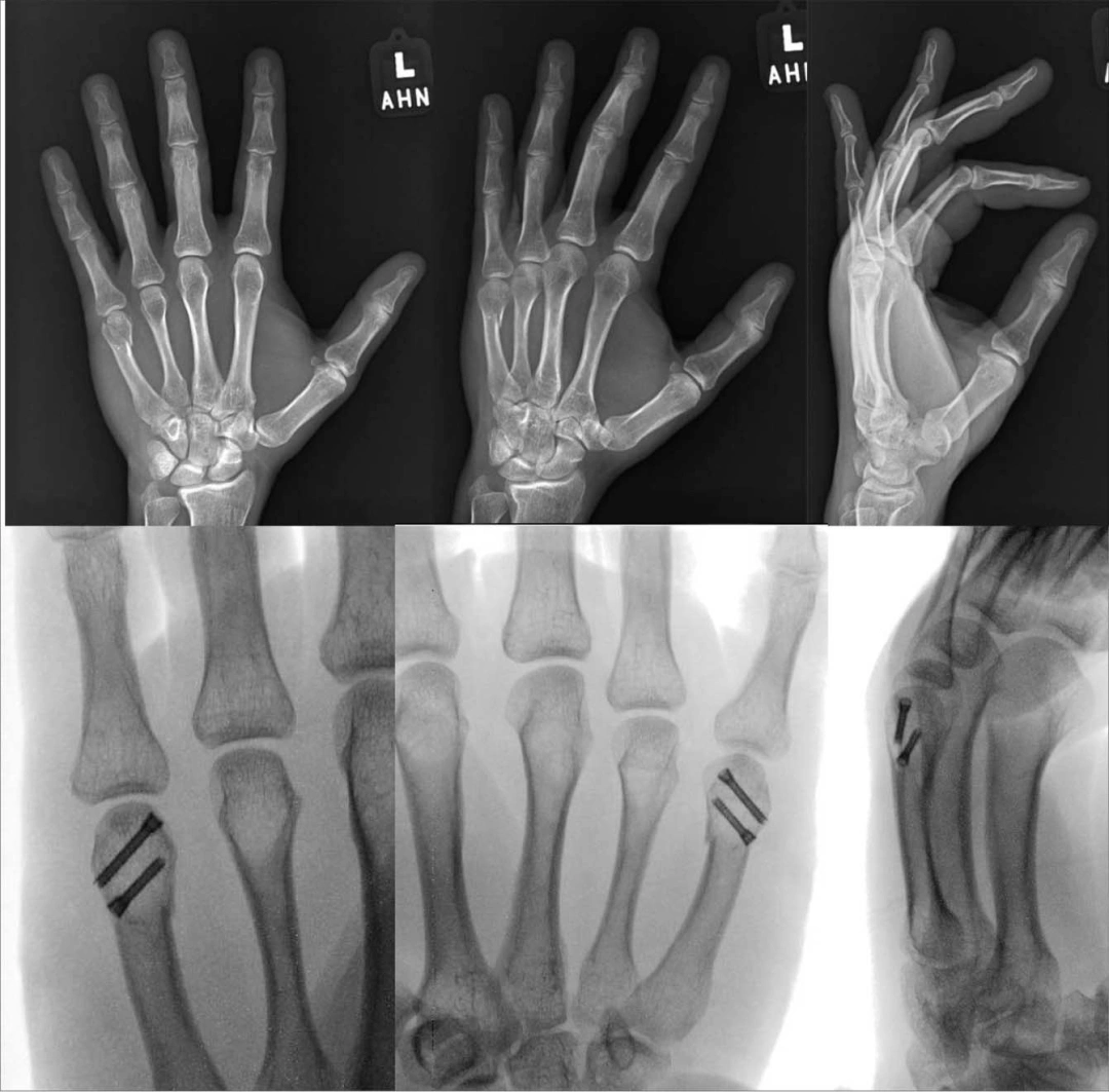
Metacarpal head fractures are less common than neck or shaft fractures. They frequently involve the articular surface and may be associated with comminution, instability, or a defect that interferes with metacarpophalangeal joint motion.
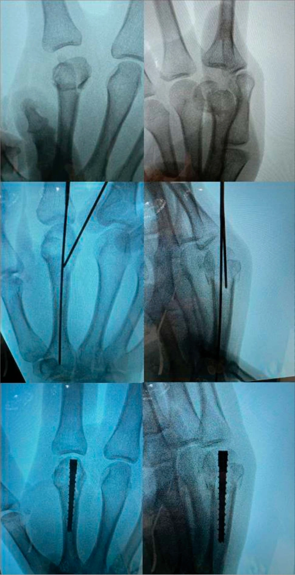
The treatment decision depends on:
The size and location of the articular fragment;
The degree of displacement;
Joint congruity;
Presence of a mechanical block;
Stability after reduction;
Amount of remaining bone available for fixation.
Small, nondisplaced fractures may be treated with immobilization and close radiographic follow-up. However, displaced intra-articular fractures or fractures that interfere with joint motion often require operative treatment.
A commonly cited surgical indication is an articular fracture with more than approximately 1 mm of displacement or a fragment that blocks joint motion. This threshold should be interpreted in the context of the complete clinical and radiographic picture rather than used as an isolated rule.
Possible fixation methods include:
Closed reduction and percutaneous K-wire fixation;
Miniature screw fixation;
Condylar plate fixation;
Combined plate and screw fixation;
Selected intramedullary fixation techniques.
For a large articular fragment, a small lag screw may provide effective compression. When the fracture extends into the metaphysis or has a more complex configuration, a mini plate may provide additional stability.
Extensively comminuted fractures may not provide enough bone stock for reliable screw purchase. In these cases, K-wire fixation may be more appropriate, although the surgeon must avoid joint penetration and excessive immobilization.
Intramedullary fixation may be considered when the distal fragment has sufficient bone stock to accommodate the implant. The technique can reduce soft-tissue dissection, but its feasibility depends heavily on:
The size of the distal fragment;
The fracture line;
The amount of comminution;
The relationship to the metacarpophalangeal joint;
The ability to obtain stable fixation without violating the articular surface.
Therefore, intramedullary fixation is not suitable for every metacarpal head fracture.
Metacarpal neck fractures are especially common in the fifth metacarpal and are often referred to as “boxer’s fractures.” They typically occur after an axial load or impact to a clenched fist.
The fracture commonly produces:
Dorsal angulation of the distal fragment;
Volar comminution;
Loss of knuckle prominence;
Pain and swelling around the metacarpophalangeal joint.
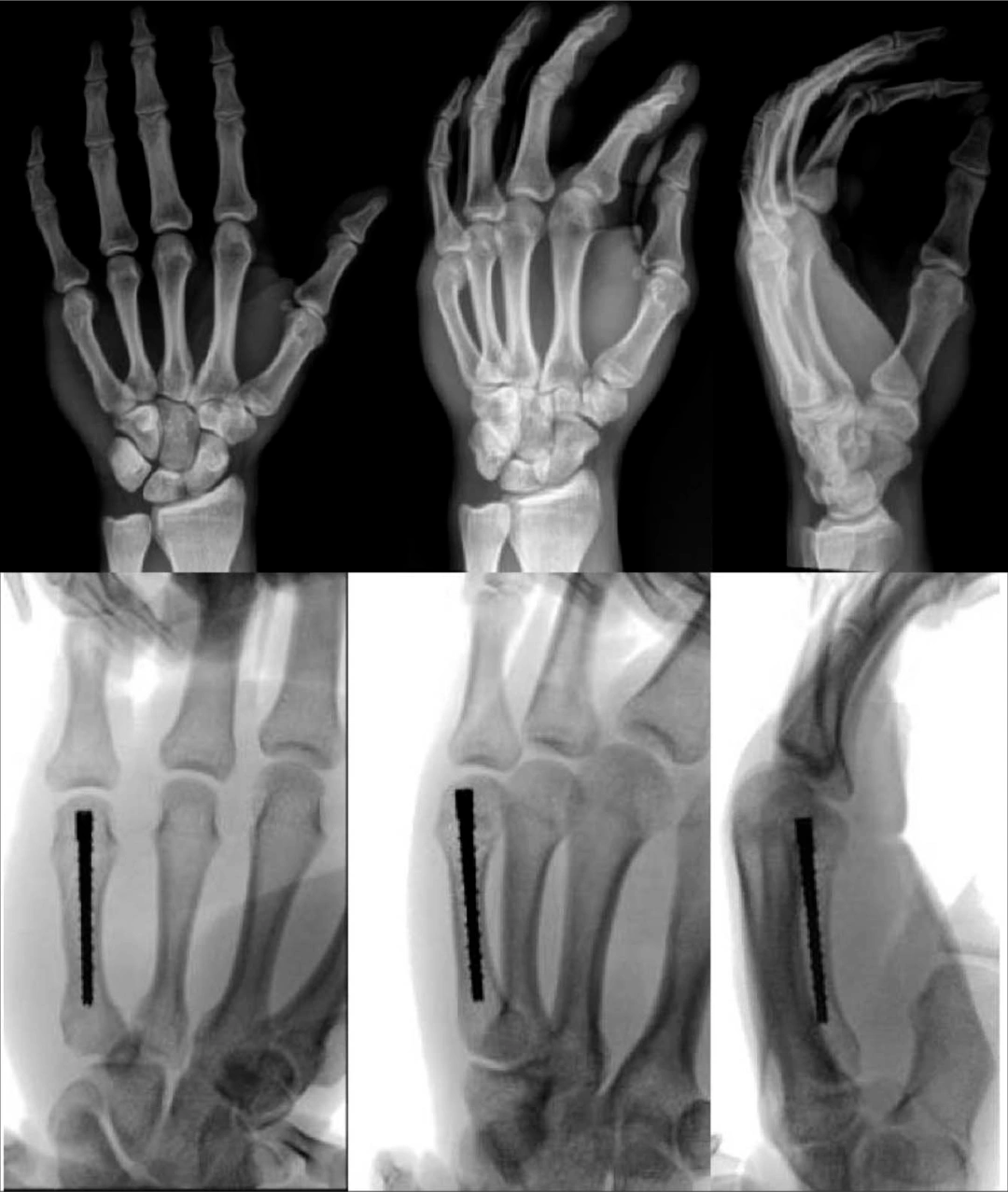
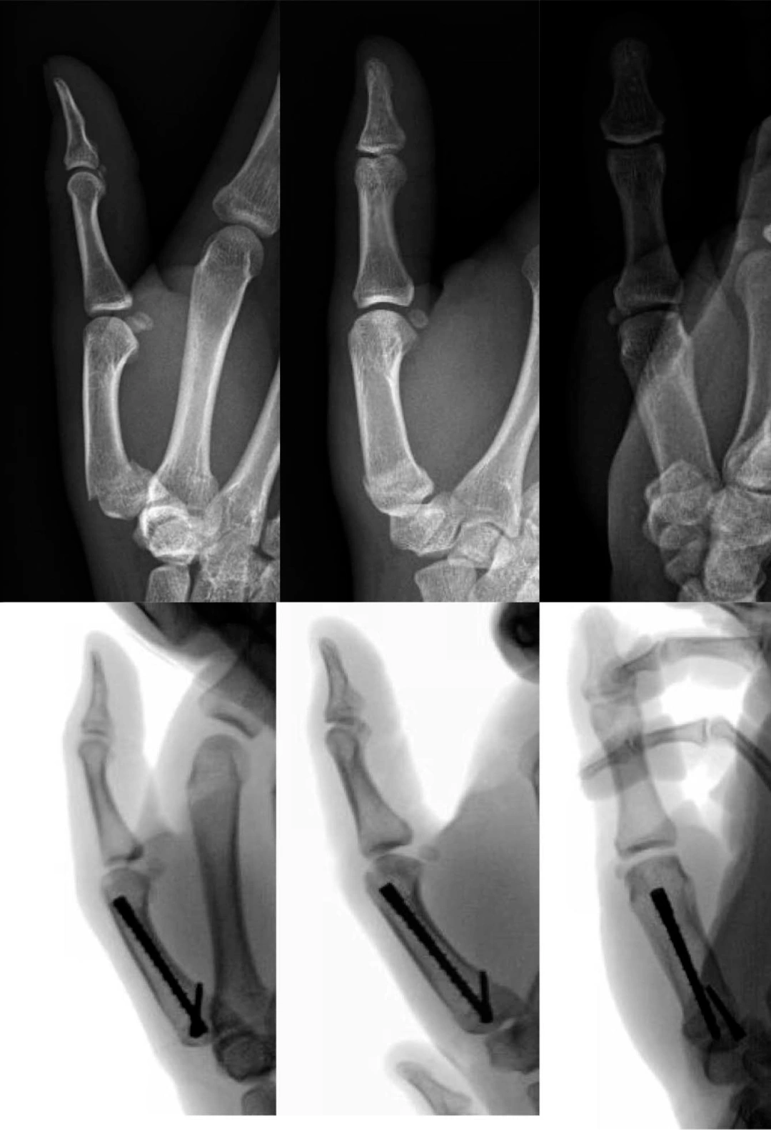
▲Intramedullary fixation is applicable to fractures with adequate bone stock.
A fifth metacarpal neck fracture without rotational deformity can often be treated successfully with immobilization, protective splinting, or buddy strapping.
The amount of acceptable angulation depends on the involved digit, the patient’s symptoms, and whether there is functional impairment. The ulnar metacarpals generally tolerate more angulation than the index and middle metacarpals, but excessive deformity may still cause weakness, extensor lag, or cosmetic problems.
The key clinical question is not simply:
“How many degrees of angulation are present?”
It is:
“Does the deformity cause unacceptable functional impairment or fail to remain stable during treatment?”
Surgical treatment may be considered for:
Open fractures;
Irreducible fractures;
Unstable fractures that redisplace;
Significant rotational deformity;
Substantial shortening;
Displaced intra-articular extension;
Multiple metacarpal fractures;
Fractures with a mechanical block to motion;
Patients who require earlier functional recovery.
There is no universally accepted optimal fixation method. A review of surgical techniques found that K-wire fixation, dorsal plating, and intramedullary fixation are all used, while complication rates and functional outcomes vary according to the technique and fracture characteristics.
K-wire fixation is widely used for unstable metacarpal neck fractures.
Possible techniques include:
Transverse pinning;
Crossed K-wire fixation;
Antegrade intramedullary pinning;
Bouquet pinning.
Minimally invasive;
Relatively simple instrumentation;
Useful when the distal fragment is small;
Suitable for selected comminuted fractures;
Usually requires less soft-tissue dissection than plating.
Pin-site infection;
Risk of loss of reduction;
Limited rotational control in some configurations;
Exposed pins may require protection;
Hardware removal may be necessary;
Prolonged immobilization can contribute to stiffness.
When the available distal bone is very limited, K-wire fixation may be preferable because it does not require multiple screws to obtain purchase in a small fragment.
Intramedullary fixation can be performed with flexible K-wires, intramedullary nails, or headless compression screws.
It may be suitable when:
The medullary canal is sufficiently large;
The distal fragment has adequate bone stock;
The fracture can be reduced satisfactorily;
Stable fixation can be achieved without damaging the joint;
Early motion is desirable.
Intramedullary fixation can reduce the need for extensive exposure and may minimize irritation from dorsal implants. However, rotational control remains an important consideration, particularly in unstable or highly comminuted fractures.
A systematic review and meta-analysis comparing intramedullary K-wires with alternative fixation methods found no consistent difference in clinical outcomes for metacarpal neck fractures. For shaft fractures, intramedullary K-wires were associated with a shorter operative time in the pooled analysis.
Plate fixation provides rigid stabilization and may be useful for unstable or comminuted fractures.
Potential advantages include:
Strong resistance to angulation;
Better control of fracture length;
Improved rotational stability;
Possibility of early controlled mobilization;
Usefulness in fractures with multiple fragments.
However, plate fixation requires greater soft-tissue exposure and may be associated with:
Extensor tendon irritation;
Adhesion;
Scar formation;
Joint stiffness;
Hardware prominence;
Need for later implant removal.
For this reason, plating should be selected when its stability benefits outweigh the potential soft-tissue disadvantages. It should not automatically be regarded as superior to less invasive fixation.
The metacarpal shaft is a common site of fracture. Shaft fractures may be transverse, oblique, spiral, or comminuted, and each pattern presents different fixation challenges.
Shaft fractures can result in:
Shortening;
Dorsal angulation;
Rotational deformity;
Extensor lag;
Reduced grip strength;
Loss of finger alignment.
Oblique and spiral fractures are particularly prone to rotation and shortening. Clinical examination during finger flexion is essential because radiographic alignment alone may not identify all rotational problems.
Surgery may be indicated for:
Open fractures;
Multiple metacarpal fractures;
Unstable fractures;
Irreducible fractures;
Loss of reduction during immobilization;
Significant shortening;
Rotational malalignment;
Displaced intra-articular extension;
Severe soft-tissue injury;
Segmental bone loss.
Commonly referenced angulation thresholds include more than approximately 10° for the index and middle metacarpals and approximately 30°–40° for the ring and small metacarpals. However, these values are not absolute. The decision must also consider rotation, shortening, fracture stability, and patient function.
K-wire fixation is frequently used for metacarpal shaft fractures, especially when a minimally invasive approach is desirable.
Possible configurations include:
Intramedullary K-wires;
Transverse intermetacarpal pinning;
Cross-pinning;
Combined fixation techniques.
Limited surgical exposure;
Relatively straightforward instrumentation;
Useful for selected transverse or short-oblique fractures;
Can preserve soft-tissue structures.
Less rigid than plate fixation in some fracture patterns;
Rotational stability may be limited;
Pin-site infection;
Potential loss of reduction;
Possible need for secondary removal.
Intramedullary K-wires can help maintain length and alignment, while transverse intermetacarpal wires may improve rotational stability in selected cases.
Interfragmentary screws may be used for long oblique or spiral fractures when the fragments are sufficiently large and the fracture geometry allows compression.
The technique can provide:
Direct interfragmentary compression;
Minimal implant prominence;
Good control of the fracture line;
Reduced need for a large plate in selected cases.
A neutralization plate may be added when the fracture requires additional protection against bending or torsional forces.
The surgeon must ensure that the screw trajectory is appropriate and that the screw does not split the fragment or interfere with tendon gliding.
Plate fixation is useful for unstable fractures that require stronger control of:
Length;
Angulation;
Rotation;
Multiple fracture fragments.
Available constructs may include:
Compression plates;
Locking plates;
Neutralization plates;
Mini-fragment plates;
Low-profile dorsal or lateral plates.
Locking plates may be particularly useful when bone quality is reduced or when conventional screw purchase is limited. Nevertheless, plate selection should account for soft-tissue coverage and the risk of extensor tendon irritation.
The purpose of fixation is to provide sufficient stability for fracture healing while allowing safe early mobilization. Excessively bulky implants or unnecessary soft-tissue disruption may compromise this objective.
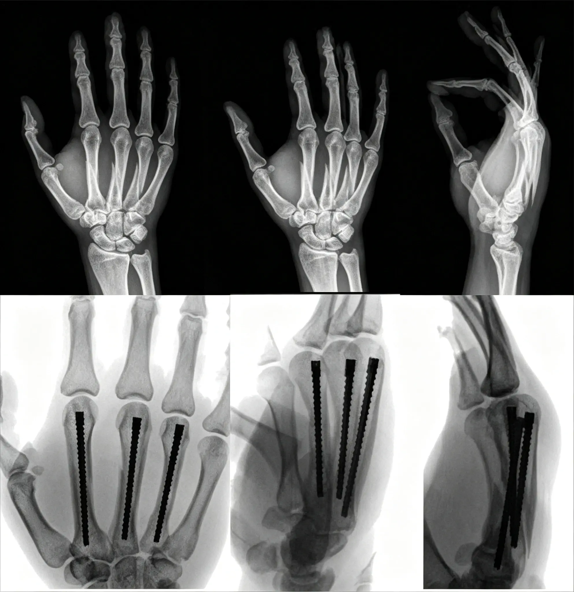
Intramedullary fixation is increasingly used for selected metacarpal shaft fractures.
Potential advantages include:
Smaller incisions;
Less periosteal stripping;
Reduced implant prominence;
Efficient fixation of selected fracture patterns;
Potentially shorter operative time.
Intramedullary fixation may be considered for certain transverse, short-oblique, spiral, or comminuted fractures, provided adequate reduction and rotational control can be achieved.
However, it is not automatically appropriate for every fracture. Important considerations include:
Canal diameter;
Fracture length;
Comminution;
Bone stock;
Implant entry point;
Rotational stability;
Risk of joint or tendon irritation.
Recent reviews support intramedullary fixation as a useful option, but available comparative evidence does not establish one technique as universally superior.
External fixation is less commonly used for routine closed metacarpal fractures. It has an important role in complex injuries involving severe soft-tissue compromise.
Potential indications include:
Open fractures;
Extensive soft-tissue defects;
Contaminated wounds;
Osteomyelitis;
Segmental bone loss;
Severe comminution;
Injuries requiring staged reconstruction.
External fixation can stabilize the bone while allowing access to the soft tissues for wound care, flap coverage, or further reconstruction.
Its limitations include:
Pin-site infection;
Bulky external components;
Patient discomfort;
Need for careful pin placement;
Potential difficulty with rehabilitation.
For uncomplicated closed fractures, internal fixation or conservative care is usually more appropriate.
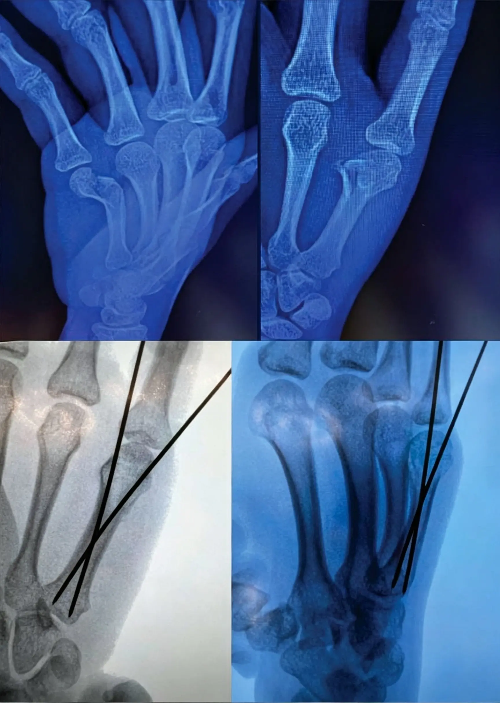
Metacarpal base fractures vary considerably depending on the involved digit and whether the carpometacarpal joint is affected.
The first metacarpal base is particularly important because it forms the thumb carpometacarpal joint, which is essential for opposition, pinch, and grasp.
Some fractures at the base of the first metacarpal can tolerate nonoperative treatment because the thumb carpometacarpal joint may compensate for limited deformity.
However, displaced or unstable intra-articular fractures may require fixation to restore:
Joint congruity;
Metacarpal alignment;
Thumb length;
Pinch strength;
Long-term joint function.
Depending on the fracture pattern, options may include:
Closed reduction and percutaneous K-wire fixation;
Interfragmentary screw fixation;
Mini-plate and screw fixation;
Intramedullary fixation;
Open reduction and internal fixation.
The choice depends on whether the fracture is:
Extra-articular;
Intra-articular;
Comminuted;
Unstable;
Associated with carpometacarpal subluxation or dislocation.
For unstable displaced fractures, restoration of the articular surface and stable alignment is generally the main priority.
The fixation method should support a rehabilitation plan rather than determine it independently.
Postoperative management may include:
Short-term immobilization;
Elevation and edema control;
Wound and pin-site care;
Serial radiographs;
Early controlled finger motion;
Progressive strengthening after clinical and radiographic healing.
The timing of mobilization depends on:
Fracture stability;
Fixation strength;
Soft-tissue condition;
Bone quality;
Presence of associated injuries;
Surgeon preference and rehabilitation protocol.
Early motion is important because prolonged immobilization may cause:
Metacarpophalangeal stiffness;
Tendon adhesions;
Reduced grip strength;
Loss of functional range of motion.
At the same time, premature loading may lead to loss of reduction, implant failure, or delayed union. Rehabilitation should therefore be progressive and guided by clinical and radiographic findings.
Complications may occur after either conservative or surgical treatment.
Loss of fracture reduction;
Malunion;
Shortening;
Rotational deformity;
Persistent pain;
Reduced grip strength;
Joint stiffness.
Infection;
Pin-site infection;
Tendon adhesion;
Extensor irritation;
Hardware prominence;
Implant failure;
Nonunion or delayed union;
Joint stiffness;
Need for implant removal;
Residual malrotation.
The risk of complications depends on the fracture pattern, soft-tissue injury, surgical approach, implant choice, and postoperative rehabilitation.
Fracture pattern | Common treatment considerations |
|---|---|
Stable, nondisplaced fracture | Splinting, cast, or protected mobilization |
Displaced metacarpal head fracture | Mini screws, K-wires, or plate fixation |
Small distal fragment | K-wire fixation may be preferable |
Unstable metacarpal neck fracture | K-wires, intramedullary fixation, or plate and screws |
Long oblique or spiral shaft fracture | Interfragmentary screws with or without a neutralization plate |
Unstable transverse shaft fracture | Plate fixation, K-wires, or intramedullary fixation |
Comminuted shaft fracture | Plate, intramedullary fixation, or selected external fixation |
Open fracture with soft-tissue loss | Debridement and staged or external fixation |
Unstable first metacarpal base fracture | Percutaneous fixation or ORIF |
Segmental bone loss or osteomyelitis | External fixation or staged reconstruction |
This table is a general overview. Actual treatment must be based on clinical examination, imaging, fracture stability, and the surgeon’s assessment.
No. Many stable fractures can be managed conservatively, but open fractures, unstable fractures, significant rotational deformity, displaced intra-articular fractures, and fractures that lose alignment may require surgery.
K-wire fixation, plate and screw fixation, interfragmentary screws, and intramedullary fixation are all commonly used. The best method depends on the fracture location and pattern.
They may be suitable for selected comminuted fractures, but the surgeon must confirm that adequate reduction and rotational stability can be achieved. Severe comminution or inadequate bone stock may favor another fixation method.
A mini plate may be preferred when a fracture is unstable, displaced, comminuted, or requires stronger control of length and rotation. However, the risk of tendon irritation and stiffness must also be considered.
Healing time varies according to fracture pattern, patient factors, and treatment. Clinical healing and return to unrestricted activity may occur at different times, and radiographic union does not always mean that the hand is ready for heavy loading.
Rotational deformity can cause the fingers to overlap or scissor when the hand is closed. This may significantly impair grip and fine motor function even when the fracture appears acceptable on radiographs.
Metacarpal fracture treatment requires a balance between fracture stability, soft-tissue preservation, and early functional recovery.
Stable fractures with acceptable alignment can often be treated with immobilization and controlled mobilization. Unstable, displaced, open, intra-articular, or rotationally malaligned fractures may require fixation using K-wires, interfragmentary screws, mini plates, intramedullary implants, or external fixation.
The most important principles are:
Correct rotational deformity;
Preserve metacarpal length and alignment;
Restore joint congruity when necessary;
Select fixation according to fracture anatomy;
Minimize unnecessary soft-tissue damage;
Begin safe rehabilitation as early as stability allows.
No single implant is ideal for every metacarpal fracture. Successful treatment depends on matching the fixation strategy to the fracture pattern and the patient’s functional requirements.