
Views: 0 Author: Site Editor Publish Time: 2026-09-20 Origin: Site
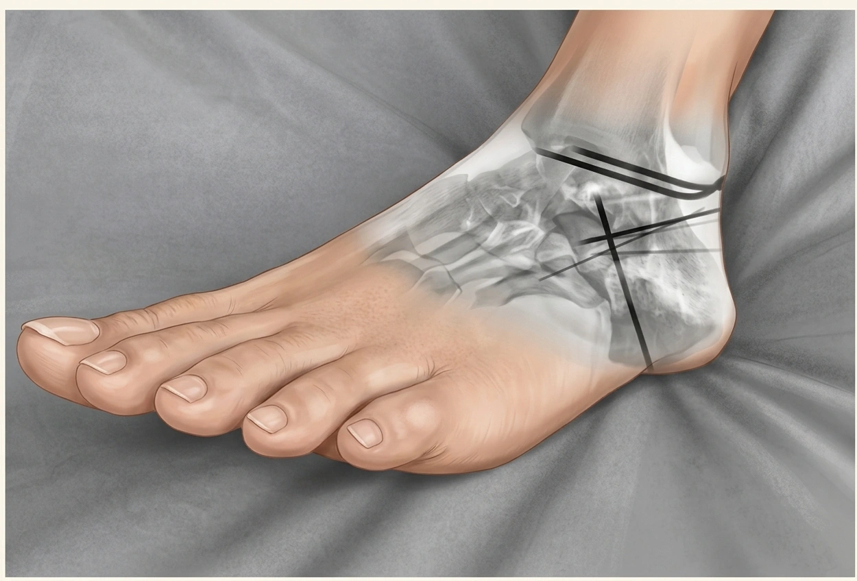
Calcaneal fractures are among the most complex injuries of the hindfoot. They are frequently caused by high-energy trauma, including falls from height and motor-vehicle accidents, and a substantial proportion involve the subtalar joint. Because of the complex three-dimensional anatomy of the calcaneus, displaced intra-articular fractures can lead to loss of hindfoot alignment, subtalar stiffness, chronic pain, altered biomechanics, and post-traumatic arthritis if the anatomy is not adequately restored.
For surgically treated displaced intra-articular calcaneal fractures, the principal objectives are to restore the congruity of the posterior facet, recover calcaneal height, length and alignment, correct varus or valgus deformity, and obtain stable fixation that allows appropriate postoperative rehabilitation.
Modern calcaneal fracture surgery increasingly emphasizes fracture-specific surgical approaches, controlled reduction, preservation of the soft-tissue envelope, and minimally invasive fixation when appropriate. Evidence comparing the extensile lateral approach (ELA) with the sinus tarsi approach (STA) suggests that both can achieve satisfactory radiographic restoration, while STA generally has a lower rate of wound complications. Therefore, approach selection should be individualized according to fracture morphology, soft-tissue condition, patient factors, and surgeon experience.
This article summarizes key operative principles and step-by-step reduction and fixation techniques described in German foot and ankle trauma practice, including the extensile lateral approach, sinus tarsi approach, medial approach, reconstruction of the posterior facet, Essex-Lopresti reduction, and minimally invasive intramedullary fixation.
The calcaneus is a three-dimensional bone that forms the foundation of the hindfoot. Its posterior facet articulates with the talus to form the subtalar joint, while the anterior process participates in the calcaneocuboid joint.
The main goals of surgical treatment are:
Restore the posterior facet of the subtalar joint
Restore calcaneal height and length
Correct calcaneal varus or valgus
Restore the normal Böhler angle
Reconstruct the angle of Gissane
Restore the lateral wall contour
Correct displacement of the anterior process
Achieve stable fixation
Minimize soft-tissue injury
Preserve or restore hindfoot biomechanics
Böhler angle and Gissane angle are useful intraoperative radiographic references, but they should not be interpreted in isolation. Fluoroscopic assessment should also evaluate overall hindfoot alignment, calcaneal height, width, length, and posterior facet congruity.
For complex fractures, CT imaging—particularly multiplanar and three-dimensional reconstruction—is valuable for understanding fracture morphology and planning the surgical approach.
Three principal approaches are commonly used for operative treatment of calcaneal fractures:
Extensile lateral L-shaped approach
Sinus tarsi approach
Medial approach
The choice should be based on fracture pattern and soft-tissue condition rather than using one approach for every fracture.
Recent evidence indicates that sinus tarsi fixation can provide similar radiographic outcomes to the extensile lateral approach in appropriately selected displaced intra-articular fractures, while reducing wound-related complications. A 2024 meta-analysis of 21 comparative studies involving 2,086 patients found a higher risk of postoperative wound complications with the extensile lateral approach than with the sinus tarsi approach.
The extensile lateral approach provides broad exposure of the lateral calcaneus and subtalar joint and remains an important option for complex displaced intra-articular fractures requiring extensive visualization and reconstruction.
The incision is generally L-shaped. The vertical limb is positioned along the posterolateral hindfoot, while the horizontal limb extends anteriorly along the lateral border of the calcaneus toward the base of the fifth metatarsal.
Careful soft-tissue handling is critical because the lateral calcaneal skin has relatively limited tolerance for surgical trauma.
After protecting the sural nerve, the soft-tissue flap is elevated as a full-thickness flap directly on the bone.
The calcaneofibular ligament can be released when necessary to improve exposure of the subtalar joint.
The peroneal tendon complex is mobilized from the lateral wall and protected within the soft-tissue flap. Attention should be paid to the structures located around the peroneal tendons and the lateral hindfoot.
Once the subtalar joint and sinus tarsi are exposed, the soft-tissue flap can be carefully retracted dorsally. Temporary fixation pins may be placed into the talar neck or other safe bony areas to maintain exposure.
This approach can provide visualization of:
The lateral calcaneal wall
Posterior facet of the subtalar joint
Sinus tarsi
Calcaneal tuberosity
Lateral talus
Major intra-articular fracture fragments
However, its extensive soft-tissue dissection is associated with a greater risk of wound complications than less invasive approaches.
The sinus tarsi approach is a limited lateral approach designed to provide direct access to the posterior facet and subtalar joint while minimizing disruption of the lateral soft tissues.
It is particularly useful for selected Sanders type II fractures and some Sanders type III fractures, although more complex fracture patterns may also be treated through minimally invasive approaches when adequate reduction can be achieved.
A typical incision is approximately 3–4 cm long and extends from the region of the lateral malleolus toward the base of the fourth metatarsal.
After opening the subcutaneous tissues, the relevant cutaneous nerve branches are identified and protected. The inferior extensor retinaculum and peroneal tendons are carefully mobilized as required.
The subtalar joint capsule is opened to expose the anterior and lateral portions of the posterior facet.
The approach can be extended slightly dorsally or distally according to fracture morphology and implant requirements.
Smaller incision
Reduced soft-tissue disruption
Direct visualization of the posterior facet
Suitable for minimally invasive reduction and fixation
Potentially lower wound complication rate
Shorter operative time in many comparative studies
Multiple meta-analyses have reported lower wound complication rates with the sinus tarsi approach compared with the extensile lateral approach, while radiographic restoration and functional outcomes are often broadly comparable.
Importantly, the sinus tarsi approach should not be regarded as universally superior. Highly comminuted fractures requiring extensive exposure may still require a larger approach or alternative fixation strategy.
The medial approach is used less frequently and is primarily considered for fractures involving medial structures, particularly displaced fractures of the sustentaculum tali.
The incision follows the medial contour of the calcaneus, extending around the sustentaculum tali region toward the plantar-medial aspect of the foot.
The tibial neurovascular structures must be carefully identified and protected. The flexor hallucis longus and flexor digitorum longus tendons can be retracted as necessary to expose the medial calcaneus and sustentaculum tali.
This approach may provide direct access to:
Sustentaculum tali
Medial wall of the calcaneus
Medial portion of the subtalar joint
Selected medial fracture fragments
Because the posterior tibial neurovascular structures are located in close proximity, meticulous anatomical dissection is essential.
Once the appropriate approach has been selected, reduction should proceed in a controlled sequence.
The fundamental principle is:
Restore the constant medial fragment and posterior facet first, then reconstruct the remaining calcaneal architecture.
The sustentaculum tali fragment is often referred to as the “constant fragment” because its ligamentous relationship with the talus tends to maintain a relatively stable anatomical position.
A published operative technique emphasizes reconstruction of the posterior facet beginning with reduction of the most medial fragment to the sustentaculum tali. The remaining fracture fragments should not be definitively reduced until the medial calcaneal wall has been restored.
The posterior calcaneal tuberosity is frequently displaced, shortened, impacted, and rotated.
The first objective is to release the impaction and restore the height and overall alignment of the calcaneus.
A percutaneous Schanz pin or Steinmann-type pin can be inserted into the tuberosity and used as a joystick.
Controlled manipulation can then be used to:
Restore calcaneal height
Correct varus or valgus
Correct shortening
Release impacted fragments
Restore the relationship between the tuberosity and posterior facet
The Böhler angle can provide a useful radiographic reference during this stage.
After reduction, the tuberosity can be temporarily fixed to the relatively stable medial fragment with K-wires.
A reduction distractor can also be useful.
Pins may be placed in the lateral talus and calcaneal tuberosity, allowing controlled three-dimensional distraction and correction.
The distractor can help:
Restore calcaneal height
Correct hindfoot deformity
Open impacted fracture lines
Maintain the reduction while the posterior facet is reconstructed
This can be particularly useful when manual manipulation alone cannot adequately restore the calcaneal architecture.
The posterior facet is the key articular component of most displaced intra-articular calcaneal fractures.
The thin lateral wall can be carefully elevated like a hinged door to improve visualization of the subtalar joint.
The sustentaculum tali or constant fragment should serve as the primary reference for posterior facet reconstruction.
If the constant fragment is displaced from the talus, it should first be reduced and temporarily stabilized.
The remaining posterior facet fragments are then reconstructed sequentially, generally from medial to lateral.
Fine bone hooks and elevators can be used to assess:
Articular surface congruity
Fragment height
Rotation
Medial-lateral alignment
Subchondral support
Temporary fixation can be achieved with approximately 1.4–1.6 mm K-wires.
Reduction should be checked using multiple fluoroscopic views.
Useful for assessing:
Böhler angle
Calcaneal height
Posterior facet alignment
Overall length
Useful for evaluating:
Hindfoot varus/valgus
Calcaneal width
Lateral wall restoration
Intraoperative fluoroscopy should be used critically before definitive fixation. Residual articular malreduction or hindfoot malalignment should be corrected before the final implant is applied.
In highly comminuted Sanders III or IV fractures, the posterior facet may be divided into multiple central fragments.
A controlled medial-to-lateral reconstruction strategy can be used when the fragment morphology allows it.
Temporary K-wire fixation should be placed carefully to avoid interfering with definitive implants.
In selected complex fractures, some posterior facet fragments can be temporarily removed, reconstructed on the sterile field, and then reinserted as a reconstructed articular unit.
This technique may be useful when the fracture configuration prevents adequate visualization or manipulation inside the wound.
However, it is technically demanding and should be reserved for appropriately selected fractures.
The objective is to create a stable reconstructed posterior facet that can then be accurately positioned against the constant fragment and talar articular surface.
In selected cases, small K-wire segments can be intentionally left buried beneath the articular surface to stabilize reconstructed fragments.
This technique has been described as a “lost K-wire” or buried K-wire technique.
When used, the wire should be positioned so that it does not protrude into the subtalar joint or interfere with later fixation.
A predetermined bending or break point can be created before insertion, allowing the wire to be broken flush with the bone surface after the desired fixation has been achieved.
Because buried K-wire fragments can be difficult to remove and may require additional bone work, this technique should not be considered routine. Implant selection should always consider the possibility of future hardware removal.
Tongue-type calcaneal fractures present a distinct reduction challenge because the Achilles tendon pulls on the posterior tuberosity fragment.
This can maintain displacement of the posterior facet and make anatomical reduction difficult.
The Essex-Lopresti maneuver uses percutaneous pins as a joystick to manipulate the tongue-type fragment.
The basic concept is:
Insert a robust pin into the tongue-type fragment.
Use the pin as a lever.
Rotate the fragment distally.
Restore the posterior facet.
Correct calcaneal height and alignment.
Temporarily stabilize the reduction.
Confirm the reduction fluoroscopically.
Perform definitive fixation.
The Essex-Lopresti technique has been described particularly for selected tongue-type fractures, including Sanders II patterns. Published clinical series have demonstrated that percutaneous reduction can achieve acceptable restoration in appropriately selected cases.
If indirect reduction cannot achieve satisfactory articular alignment, conversion to an open technique may be necessary. Treatment should therefore be individualized rather than forcing a minimally invasive technique when reduction quality is inadequate.
The anterior process of the calcaneus should also be assessed after reconstruction of the posterior facet.
This is particularly important when the fracture extends into the calcaneocuboid joint.
The anterior process should be restored anatomically to re-establish:
Calcaneocuboid joint congruity
Calcaneal length
The transition between the anterior process and the angle of Gissane
Overall lateral column alignment
The angle of Gissane is a useful radiographic landmark for evaluating the central architecture of the calcaneus.
After reduction, temporary K-wire fixation can be used before definitive fixation.
A key principle is:
Definitive fixation should begin only after all clinically relevant fracture fragments have been satisfactorily reduced.
Once reduction has been confirmed, definitive fixation can be selected according to fragment size, fracture morphology, bone quality, surgical approach, and soft-tissue condition.
Approximately 2.0–3.5 mm screws may be used for smaller articular fragments when appropriate.
The screw trajectory should avoid penetrating the subtalar joint or calcaneocuboid joint.
Lateral locking plates remain an important fixation option for displaced intra-articular calcaneal fractures treated through open or limited lateral approaches.
A plate can provide:
Lateral wall support
Multiple fixation points
Subchondral support
Stable fixation of reconstructed fragments
Control of calcaneal width and alignment
Before plate placement, fluoroscopy should confirm restoration of calcaneal height, width, alignment, and posterior facet congruity.
Minimally invasive intramedullary fixation has become another option for selected displaced intra-articular calcaneal fractures.
The Calcanail® technique uses a plantar entry point and an intramedullary fixation concept that can combine reduction and fixation through a relatively limited surgical corridor.
After establishing the working channel, reduction instruments can be used to restore:
Calcaneal height
Calcaneal length
Hindfoot alignment
Posterior facet position
Additional sinus tarsi access may be used when direct visualization or manipulation of the posterior facet is necessary.
The nail is then locked at multiple levels to stabilize the reconstructed calcaneus.
Published clinical studies have reported acceptable radiographic and functional outcomes with Calcanail® in selected Sanders II and III fractures.
A recent randomized controlled trial also reported favorable perioperative and 24-month outcomes with a minimally invasive intramedullary nail technique compared with extensile lateral plate fixation in selected Sanders II–III fractures, although these results should not be generalized to every calcaneal fracture pattern or every nail system.
Another minimally invasive option is the C-Nail® system.
The technique may begin with a sinus tarsi approach to reconstruct the posterior facet and restore the overall calcaneal anatomy.
After reduction, an intramedullary nail is introduced through the posterior calcaneal region toward the central calcaneal axis and secured using multiple locking elements.
Clinical reports from German trauma centers have described the use of C-Nail® fixation for severe calcaneal fractures, including Sanders III and IV injuries.
The potential advantages of intramedullary fixation include:
Smaller surgical exposure
Reduced disruption of the lateral soft tissues
Stable internal fixation
Ability to combine reduction and fixation through minimally invasive techniques
However, nail fixation requires careful fracture selection and accurate three-dimensional reduction. Severe comminution may still require alternative fixation strategies.
There is no single surgical approach that is appropriate for every calcaneal fracture.
| Fracture / Clinical Situation | Potential Strategy |
|---|---|
| Sanders II, relatively simple displacement | Sinus tarsi approach + screw/plate fixation |
| Selected Sanders III | Sinus tarsi or limited approach with direct/indirect reduction |
| Complex comminuted intra-articular fracture | Extensile lateral approach or selected minimally invasive strategy |
| Tongue-type fracture | Essex-Lopresti/percutaneous reduction when feasible |
| Medial sustentaculum tali fracture | Medial approach when direct access is required |
| Poor soft-tissue condition | Delayed, percutaneous, or minimally invasive fixation when appropriate |
| Selected Sanders II–III fractures | Intramedullary calcaneal nail |
| Severe comminution / reconstruction failure | Individualized fixation or salvage strategy |
The current literature supports a more individualized approach rather than a universal “best” technique. In particular, minimally invasive approaches can reduce wound morbidity, while extensive exposure may still be necessary when fracture complexity prevents reliable reduction through a limited incision.
Several technical errors can compromise the outcome of calcaneal fracture surgery.
The posterior facet should be reconstructed around the stable sustentaculum tali fragment whenever possible.
Restoration of hindfoot alignment is essential. Axial imaging should be reviewed before final fixation.
A visually acceptable lateral radiograph does not necessarily guarantee anatomical articular congruity. Broden views, CT, or arthroscopic assessment may be useful in selected cases.
Failure to restore calcaneal width can cause postoperative shoe-fitting problems and peroneal tendon irritation.
Every definitive screw should be checked fluoroscopically in multiple planes.
Soft-tissue preservation remains one of the most important considerations in calcaneal fracture surgery.
Minimally invasive surgery should not come at the expense of inadequate reduction. If satisfactory reduction cannot be achieved, a more extensile approach may be necessary.
Postoperative management depends on fracture stability, fixation quality, bone quality, soft-tissue status, and associated injuries.
Typical principles include:
Elevation and soft-tissue monitoring
Protection of the surgical wound
Early assessment of neurovascular status
Progressive ankle and subtalar range-of-motion exercises when fixation permits
Delayed weight bearing for fractures requiring protected healing
Serial radiographs to evaluate maintenance of reduction
Gradual progression toward partial and full weight bearing according to healing
Rehabilitation should be individualized rather than based solely on the implant type.
Potential complications include:
Wound dehiscence
Surgical-site infection
Skin necrosis
Sural nerve irritation or injury
Peroneal tendon irritation
Malunion
Loss of reduction
Subtalar stiffness
Post-traumatic subtalar arthritis
Calcaneocuboid joint degeneration
Hardware irritation
Need for implant removal
Persistent hindfoot pain
The risk profile varies according to surgical approach and patient factors. Recent evidence consistently shows greater wound-related risk with extensile lateral exposure compared with sinus tarsi techniques, although wound complications can still occur after minimally invasive surgery. A 2026 multicenter study reported wound complications in 9% of fractures treated through a sinus tarsi approach, with only one case requiring surgical revision.
The main goals are restoration of the posterior facet, calcaneal height and length, hindfoot alignment, and stable fixation while minimizing additional soft-tissue injury.
Current comparative evidence shows that the sinus tarsi approach generally has fewer wound complications and shorter operative times, while both approaches can achieve comparable radiographic restoration in appropriately selected fractures. The choice should depend on fracture morphology and soft-tissue conditions rather than a universal preference.
Sanders classification is based on the number and location of fracture lines involving the posterior facet on coronal CT images. Sanders II fractures generally involve two major articular fragments and are commonly considered candidates for operative reconstruction when displacement and clinical factors warrant surgery.
The sustentaculum tali fragment is commonly called the constant fragment because its ligamentous relationship with the talus tends to maintain its position relative to the talus. It can therefore serve as an important reference during posterior facet reconstruction.
It is particularly useful for selected tongue-type calcaneal fractures in which the posterior facet and tuberosity fragment can be manipulated indirectly using percutaneous pins.
Yes. Intramedullary calcaneal nails are an option for selected displaced intra-articular fractures, particularly when a minimally invasive strategy is appropriate. Clinical studies have reported acceptable results with systems such as Calcanail® and C-Nail®.
CT with multiplanar reconstruction is particularly useful for defining intra-articular fracture morphology. Three-dimensional reconstruction can also assist surgical planning in complex fractures.
Successful calcaneal fracture surgery depends less on a single implant or surgical approach than on a systematic understanding of fracture morphology, soft-tissue condition, and three-dimensional hindfoot anatomy.
The essential principles are to:
Select the approach according to fracture morphology and soft-tissue status.
Restore the constant medial fragment and posterior facet.
Re-establish calcaneal height, length, width, and hindfoot alignment.
Use Böhler and Gissane angles as radiographic references rather than isolated endpoints.
Confirm articular congruity with appropriate fluoroscopic views or arthroscopy when indicated.
Use temporary K-wire fixation to maintain reduction before definitive fixation.
Select plates, screws, or intramedullary nails according to fracture configuration and fixation requirements.
Avoid excessive soft-tissue disruption.
Convert to a more extensile technique when minimally invasive reduction is inadequate.
For appropriately selected fractures, modern sinus tarsi, percutaneous, and intramedullary techniques can reduce soft-tissue morbidity while maintaining the goals of anatomical reconstruction. At the same time, extensive lateral exposure remains an important tool for fractures that require broad visualization and complex reconstruction. Current evidence therefore supports a fracture-specific and patient-specific surgical strategy rather than a one-size-fits-all approach.


