
Views: 0 Author: Site Editor Publish Time: 2026-09-11 Origin: Site
Why the Iliolumbar Ligament Matters in Low Back and Hip Function
The iliolumbar ligament is one of the deepest and most clinically interesting ligamentous structures in the lumbopelvic region. Although it is relatively small compared with major muscles such as the quadratus lumborum or erector spinae, its anatomical position places it at an important junction between the lumbar spine, pelvis, sacroiliac region, thoracolumbar fascia, and hip.
Because of its deep location, the iliolumbar ligament is easy to overlook during routine physical examination. Yet anatomical and biomechanical studies suggest that it contributes to stabilization of the lumbosacral junction and may participate in the transmission of forces between the lumbar spine and pelvis.
Understanding its anatomy may therefore help clinicians better appreciate certain patterns of low back pain, lumbopelvic instability, and impaired trunk-to-hip force transfer.
However, it is important to distinguish established anatomy from biomechanical hypotheses. The iliolumbar ligament is certainly an important stabilizing structure, but claims that it directly "stores" abdominal pressure or functions like a muscle should be interpreted cautiously.
The iliolumbar ligament is a strong connective-tissue structure located at the transition between the lower lumbar spine and the pelvis.
The name itself describes its anatomical relationship:
Ilio- refers to the ilium;
Lumbar refers to the lumbar spine.
The ligament is generally described as extending from the transverse/costal process of the fifth lumbar vertebra (L5) toward the iliac crest and adjacent iliac structures.
Its anatomy is more variable than the simplified textbook description suggests. Different studies have described one, two, or multiple bands, and the morphology can vary considerably between individuals.
A recent anatomical study of 60 iliolumbar ligaments from 30 cadavers found that approximately 73% were single-banded, about 22% were double-banded, and a smaller proportion demonstrated more complex configurations. The mean ligament length was approximately 31.7 mm. Importantly, the study found no convincing evidence of a true, biomechanically meaningful insertion onto L4.
This is an important distinction when discussing iliolumbar ligament anatomy.
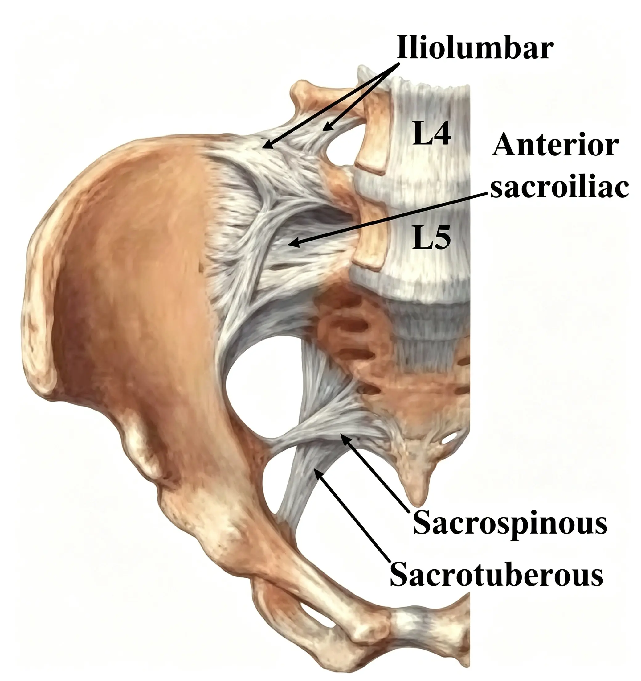
The iliolumbar ligament lies deep within the lumbopelvic region.
From a posterior approach, it is difficult to visualize directly because it is covered by multiple layers of soft tissue, including:
Skin;
Subcutaneous tissue;
Deep fascia;
Thoracolumbar fascia;
Paraspinal musculature;
Quadratus lumborum and other deep structures.
Its deep anatomical position explains why direct palpation of the ligament itself is difficult.
Imaging studies have demonstrated considerable variation in its orientation and morphology. One MRI study described two principal components arising from the L5 transverse process:
An anterior band, which is broader and flatter;
A posterior band, which is narrower and more rounded.
The anterior component extends toward the anterior portion of the iliac tuberosity, while the posterior component extends toward the iliac crest.
One of the most interesting aspects of iliolumbar ligament anatomy is that different anatomical studies have described different configurations.
Older descriptions have reported multiple bands, whereas more recent anatomical work suggests that a single-band configuration is common, with a double-band configuration representing an important anatomical variation.
The morphology may include:
Single-band structures;
Anterior and posterior bands;
Short additional collagenous bands;
Fibrous connections to surrounding fascia;
A sacroiliac component in some individuals.
The sacroiliac component is particularly interesting because anatomical studies have demonstrated fibers extending toward the sacrum and blending with the interosseous sacroiliac ligament complex. This suggests that the iliolumbar ligament may contribute not only to lumbosacral stability but also to stabilization of the sacroiliac region.
The lumbosacral junction is exposed to substantial mechanical forces during:
Standing;
Walking;
Running;
Lifting;
Bending;
Rotation;
Single-leg activities.
The iliolumbar ligament helps limit excessive movement between the lower lumbar spine and pelvis.
This stabilizing function becomes particularly important because the pelvis serves as a mechanical bridge between the spine and lower extremities.
In simple terms:
Lumbar spine → lumbosacral junction → pelvis → hip → lower extremity
The iliolumbar ligament is positioned directly within this transition zone.
Anatomical and biomechanical studies suggest that its orientation allows it to resist excessive translational and rotational movement at the lumbosacral junction.
The iliolumbar ligament does not function in isolation.
It exists within a complex network of muscles, fascia, ligaments, and connective tissues that collectively contribute to lumbopelvic stability.
The thoracolumbar fascia (TLF) is particularly important.
The thoracolumbar fascia forms several layers around the posterior abdominal wall and paraspinal muscles. It provides connective-tissue continuity between the trunk and pelvis and interacts mechanically with surrounding muscles.
Important muscles associated with this lumbopelvic system include:
Multifidus;
Erector spinae;
Transversus abdominis;
Internal oblique;
Quadratus lumborum;
Psoas major;
Iliacus.
These structures do not simply work as independent anatomical units.
Instead, they participate in coordinated control of:
Spinal stability;
Pelvic positioning;
Trunk stiffness;
Load transfer;
Hip movement.
This is one reason why evaluating only the painful structure may sometimes fail to explain persistent lumbopelvic dysfunction.
The original concept that the iliolumbar ligament can "store the kinetic energy generated by deep core muscles" is an interesting biomechanical hypothesis, but it should not be presented as an established physiological fact.
Ligaments are viscoelastic connective tissues. They can deform under load and contribute to passive mechanical resistance and elastic energy storage.
However, the iliolumbar ligament should be regarded primarily as a passive stabilizing structure, rather than an active muscle.
A more scientifically accurate model is:
Muscle contraction → fascial and skeletal force transmission → ligament loading → lumbopelvic stabilization
The ligament can therefore participate in the transmission and regulation of mechanical forces without actively generating those forces itself.
This distinction is important when discussing the relationship between the iliolumbar ligament and the deep core musculature.
The deep core system is commonly discussed in relation to spinal and pelvic stability.
Key muscles include:
The multifidus contributes to segmental control of the lumbar spine and helps regulate intervertebral movement.
The transversus abdominis contributes to abdominal wall tension and trunk stabilization.
The internal oblique participates in trunk rotation, abdominal wall tension, and coordinated trunk movement.
The erector spinae provides extension and postural control of the lumbar spine.
The quadratus lumborum contributes to lumbar stabilization, lateral flexion, and pelvic elevation.
The iliacus, together with the psoas major, is an important hip flexor and also contributes to lumbopelvic mechanics.
The interaction between these muscles and passive connective tissues is complex. The thoracolumbar fascia provides an important anatomical interface between the posterior trunk muscles and the abdominal wall musculature.
Intra-abdominal pressure (IAP) is frequently discussed as part of the body's trunk stabilization strategy.
During activities such as lifting, bracing, coughing, and rapid movement, coordinated activation of the abdominal wall, diaphragm, pelvic floor, and spinal musculature can modify intra-abdominal pressure and trunk stiffness.
However, it would be inaccurate to state that the iliolumbar ligament itself generates intra-abdominal pressure.
Instead, a more appropriate model is:
Respiratory and abdominal muscle activation
↓
Changes in intra-abdominal pressure and trunk stiffness
↓
Force transmission through the thoracolumbar and lumbopelvic connective-tissue system
↓
Improved control of the lumbar spine and pelvis
The iliolumbar ligament forms part of this passive stabilizing environment.
The relationship between the lumbar spine and hip is particularly important during walking.
During gait, the trunk and pelvis must remain sufficiently stable while the lower extremities move underneath the body.
The quadratus lumborum and iliopsoas/iliacus complex have important roles in this system.
The quadratus lumborum can contribute to pelvic elevation, sometimes described clinically as hip hiking, while the iliacus is a major contributor to hip flexion.
Therefore, dysfunction within the lumbopelvic system can potentially alter the mechanical environment in which these muscles operate.
However, this does not mean that an abnormal iliolumbar ligament is necessarily the primary cause of hip-flexor or quadratus-lumborum dysfunction.
Clinical assessment should consider the entire kinetic chain.
The iliolumbar ligament has been proposed as a potential source of pain in selected patients with low back or lumbopelvic symptoms.
Anatomical studies have identified variations in the ligament and its iliac insertion, and some authors have proposed that mechanical overload at the iliac attachment may contribute to localized pain.
The term iliolumbar syndrome has historically been used to describe pain attributed to this region.
However, iliolumbar ligament pain should be approached cautiously because low back pain is multifactorial.
Potential pain generators include:
Intervertebral discs;
Facet joints;
Sacroiliac joint;
Paraspinal muscles;
Thoracolumbar fascia;
Ligaments;
Hip pathology;
Neural structures.
Therefore, the presence of tenderness around the iliac crest does not automatically prove that the iliolumbar ligament is the source of symptoms.
Ligaments are not simply mechanical ropes.
Many ligamentous structures contain sensory nerve endings capable of providing information about tissue tension and joint position.
This has led researchers to investigate the potential sensory contribution of the iliolumbar ligament to lumbopelvic control.
However, the exact contribution of iliolumbar ligament mechanoreceptors to human posture and gait remains incompletely understood.
It is therefore more appropriate to describe the ligament as potentially contributing to proprioceptive and sensory feedback within the lumbopelvic system, rather than claiming that it independently controls posture or walking.
The iliolumbar region is anatomically close to several important nerves.
Depending on the exact anatomical location and individual variation, structures in this region may be near branches of the lumbar plexus and nerves supplying the abdominal wall and upper thigh.
For example, the:
Ilioinguinal nerve;
Iliohypogastric nerve;
Lateral femoral cutaneous nerve
may be clinically relevant when evaluating pain around the lower abdomen, groin, iliac crest, or lateral thigh.
However, these nerves should not be described as routinely lying directly "on top of" the iliolumbar ligament. Their relationship varies according to the anatomical plane and individual anatomy.
This distinction is particularly important when considering regional injections, surgical approaches, or pain procedures.
A common clinical scenario is a patient with persistent tightness or overactivity of the:
Quadratus lumborum;
Iliopsoas;
Iliacus;
Lumbar erector spinae.
It may be tempting to treat the painful or tight muscle directly.
However, persistent symptoms may sometimes reflect a broader movement-control problem.
For example:
Reduced trunk control
→ altered pelvic stabilization
→ increased demand on accessory stabilizers
→ increased quadratus lumborum or hip-flexor activity
→ altered lumbopelvic movement
→ persistent symptoms.
This does not prove that deep core weakness is the cause of every case of muscle tightness. Rather, it provides a useful framework for considering the interaction between the spine, pelvis, hip, muscles, fascia, and passive connective tissues.
When a patient presents with chronic quadratus lumborum or iliopsoas-related symptoms, treating the symptomatic muscle alone may not always be sufficient.
A comprehensive assessment can include:
Range of motion;
Segmental mobility;
Pain provocation;
Neurological examination.
Pelvic alignment;
Sacroiliac region;
Load transfer;
Single-leg control.
Abdominal wall control;
Trunk stabilization;
Breathing mechanics;
Multifidus function.
Hip flexion;
Hip extension;
Internal and external rotation;
Hip abductor strength;
Hip flexor function.
Walking;
Squatting;
Single-leg stance;
Running;
Lifting mechanics.
This broader assessment can help clinicians determine whether the apparent "tight muscle" is actually a compensatory response to another movement or stability problem.
The most useful way to understand the iliolumbar ligament is not to think of it as an isolated structure.
Instead, consider it as part of a larger lumbopelvic stabilization system.
The system includes:
Lumbar vertebrae
↓
Iliolumbar ligament
↓
Ilium and sacroiliac region
↓
Thoracolumbar fascia
↓
Deep trunk musculature
↓
Quadratus lumborum and hip muscles
↓
Lower extremity
The iliolumbar ligament provides passive mechanical restraint while the surrounding muscles provide active control.
Together, these structures help maintain stability while allowing sufficient mobility for walking, running, lifting, and other functional movements.
Several practical lessons can be drawn from the current anatomical evidence.
It should not be treated as a single identical structure in every patient. Its number of bands, orientation, and attachment patterns can vary considerably.
Modern anatomical evidence strongly supports the importance of the L5 transverse/costal process. A true L4 insertion appears to be uncommon or insufficiently supported by recent anatomical evidence.
The ligament helps restrict excessive movement at the lumbosacral junction and may also contribute to sacroiliac stabilization.
The iliolumbar ligament interacts anatomically and mechanically with the surrounding fascia and muscles rather than functioning independently.
Persistent quadratus lumborum or iliopsoas symptoms should not automatically be attributed to local muscle pathology.
Core muscle activation and intra-abdominal pressure contribute to trunk stability, but the iliolumbar ligament should be regarded as a passive stabilizing structure rather than an active generator of pressure.
It is located deep in the lumbopelvic region, connecting the lower lumbar spine—particularly the L5 transverse/costal process—with the ilium. Its posterior and anterior components have different orientations and attachment areas.
Although some traditional anatomical descriptions include L4, recent anatomical evidence does not provide convincing support for a true, biomechanically meaningful L4 insertion in typical iliolumbar ligaments. The principal vertebral attachment is associated with L5.
It may contribute to pain in selected patients, particularly around its iliac attachment, but low back pain is multifactorial. Iliolumbar ligament pathology should therefore be considered only after appropriate clinical assessment.
No. It is a ligament composed primarily of dense connective tissue. It does not actively contract like a muscle. It can, however, deform under mechanical load and participate in passive stabilization and force transmission.
Important surrounding structures include the quadratus lumborum, multifidus, erector spinae, transversus abdominis, internal oblique, psoas major, and iliacus.
It helps stabilize the transition between the lumbar spine and pelvis while the lower extremities move. This provides a stable base from which the hip and lower-limb muscles can generate movement.
The iliolumbar ligament is a small but strategically positioned structure within the human lumbopelvic system.
Modern anatomical research shows that its morphology is considerably more variable than traditional textbook descriptions suggest. The ligament is primarily associated with the L5 transverse/costal process and the iliac region, with different individuals demonstrating single-band, double-band, and other anatomical configurations.
Its importance extends beyond a simple attachment between bone structures. Together with the thoracolumbar fascia, lumbar musculature, abdominal wall, quadratus lumborum, iliopsoas, and sacroiliac ligament complex, it forms part of an integrated system responsible for lumbopelvic stability and efficient force transfer.
For clinicians, perhaps the most important lesson is this:
When chronic low-back, quadratus-lumborum, or hip-flexor symptoms persist, do not evaluate the painful muscle in isolation. Look at the entire lumbopelvic system.
Understanding the relationship between the lumbar spine, iliolumbar ligament, fascia, core musculature, pelvis, and hip can provide a more complete framework for evaluating movement dysfunction and lumbopelvic pain.


 16. Clinical Takeaways


