
Views: 0 Author: Site Editor Publish Time: 2026-10-20 Origin: Site
Introduction
Intertrochanteric femur fractures are extracapsular fractures of the proximal femur occurring between the greater and lesser trochanters. They are particularly common in older adults with osteoporosis and frequently result from low-energy falls. In younger patients, these fractures are more often associated with high-energy trauma, including falls from height and motor vehicle collisions.
Successful treatment depends on accurate fracture classification, assessment of mechanical stability, appropriate reduction, and selection of a suitable fixation device. The primary objective of treatment is to restore the biomechanical integrity of the proximal femur, achieve stable fixation when surgery is indicated, and facilitate early mobilization while minimizing complications.
Several classification systems are used in clinical practice, including the Evans classification, Boyd and Griffin classification, and AO/OTA classification. Common surgical fixation options include the dynamic hip screw (DHS), proximal femoral nail antirotation (PFNA), and INTERTAN intramedullary nail.
This article reviews the applied anatomy, injury mechanisms, clinical classification, diagnosis, treatment strategies, surgical techniques, postoperative rehabilitation, and traditional Chinese medicine approaches described in the management of intertrochanteric femur fractures.
Understanding proximal femoral anatomy is essential for evaluating fracture stability and planning internal fixation.
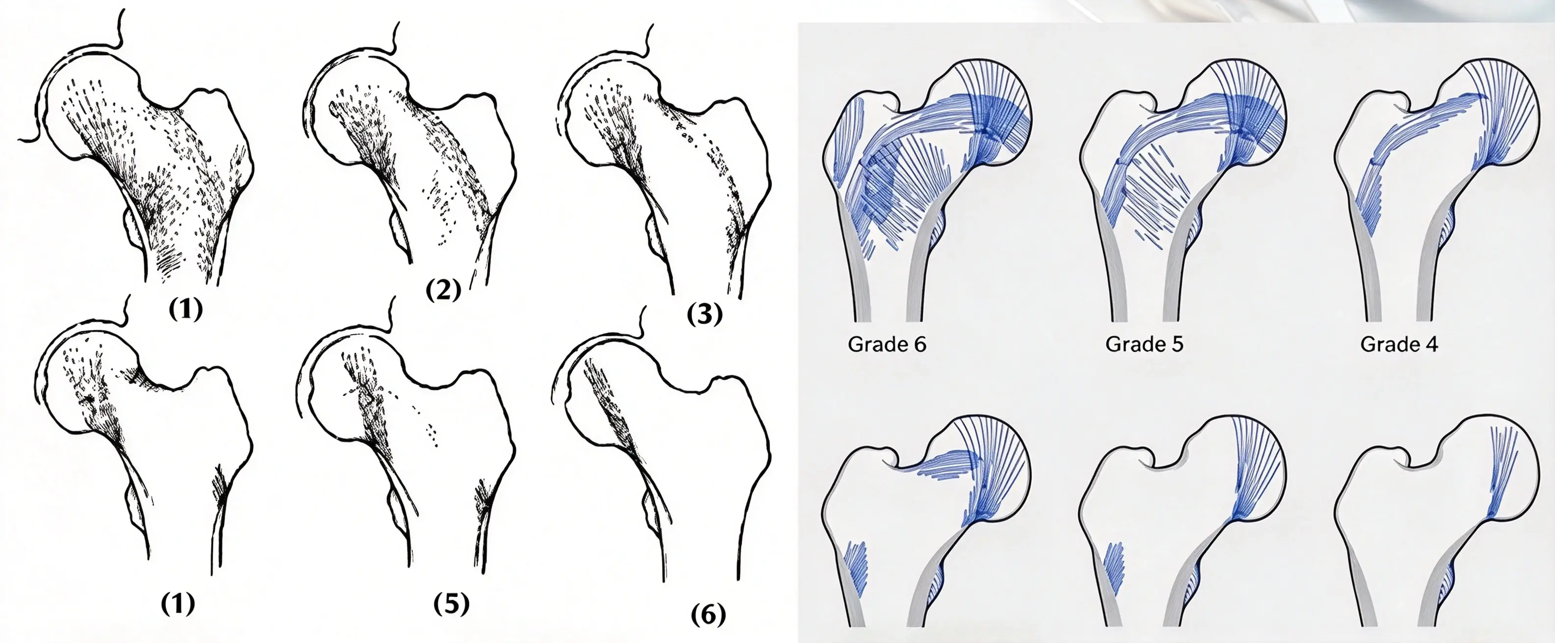
Ward's triangle is a relatively low-density region within the trabecular architecture of the proximal femur, traditionally described in the area where trabecular groups intersect within the femoral neck. Its appearance and significance vary with age, bone density, and imaging technique.
In older adults with osteoporosis, reduced trabecular bone strength can compromise the structural support available for fracture fixation.
The trabecular architecture of the proximal femur is organized to accommodate different loading directions. Changes in bone density and trabecular structure can affect the mechanical strength of the femoral neck and trochanteric region.
The calcar femorale is a dense region of bone located in the posteromedial proximal femur, extending from the region of the femoral shaft toward the femoral neck.
It contributes to load transfer and structural support within the proximal femur. The integrity of the posteromedial cortex and calcar region is important when evaluating intertrochanteric fracture stability.
When a fracture disrupts medial cortical support, maintaining reduction and fixation may become more challenging. Restoration of alignment and cortical contact, where possible, is therefore an important principle of surgical treatment.
The mechanical strength of the proximal femur depends on bone density, trabecular architecture, cortical thickness, and the distribution of physiological loads.
The original six-grade description of proximal femoral mechanical strength may be retained when supported by the accompanying anatomical illustration or source reference. However, a six-grade diagram should not be presented as a universally accepted clinical classification unless the underlying grading system is identified.
The mechanism of an intertrochanteric fracture varies according to patient age, bone quality, and the direction and magnitude of the applied force.
In younger patients, intertrochanteric fractures are commonly associated with high-energy injuries, including falls from height and motor vehicle collisions.
These injuries may involve substantial fracture displacement, comminution, and damage to surrounding soft tissues. Associated injuries must also be considered, particularly trauma involving the head, spine, chest, abdomen, or other parts of the musculoskeletal system.
A systematic trauma assessment is important before definitive fracture management.
Falls are a major cause of intertrochanteric fractures in older adults, particularly those with osteoporosis.
Factors that increase the risk of falling include:
Reduced visual acuity
Lower-limb muscle weakness
Blood pressure instability or orthostatic hypotension
Slower reaction times and impaired balance
Cardiovascular and vascular disorders
Existing bone and joint diseases
Medication-related dizziness or impaired coordination
In older adults, the combination of increased fall risk and reduced bone strength can result in a fracture after relatively minor trauma.
The likelihood of a hip fracture following a fall depends on several interacting factors:
Severity of osteoporosis: Reduced bone strength increases susceptibility to fracture.
Direction of the fall: Direct impact on the hip or the area immediately surrounding it may increase the force transmitted to the proximal femur.
Protective responses: An inadequate protective response may prevent the body from dissipating the energy of a fall.
Soft-tissue cushioning: The ability of the soft tissues around the hip to absorb impact influences the force transmitted to the bone.
Bone strength: The structural integrity of the proximal femur determines its ability to withstand the applied load.
Fall prevention and osteoporosis management are therefore important components of fracture prevention.
Direct trauma
A direct blow to the hip, such as during a motor vehicle collision or a fall onto a hard surface, may produce an intertrochanteric fracture. The resulting fracture configuration depends on the magnitude, direction, and point of impact.
Indirect trauma
Rotational forces and bending stresses can also contribute to fracture formation. Varus and anterior angulation forces may act on the proximal femur, while muscular forces influence the displacement of fracture fragments.
The lesser trochanter may become a separate fragment, and traction from the iliopsoas muscle may contribute to fragment displacement. Strong muscular forces around the greater trochanter can also affect the position of the greater trochanteric fragment.
The final fracture pattern reflects the interaction of bone strength, loading direction, and surrounding soft-tissue forces.
A clinically useful fracture classification should be straightforward to apply, support treatment planning, and provide information relevant to prognosis.
For intertrochanteric fractures, two particularly important questions are whether the fracture is mechanically stable and whether the fracture can maintain acceptable alignment under physiological loading after reduction and fixation.
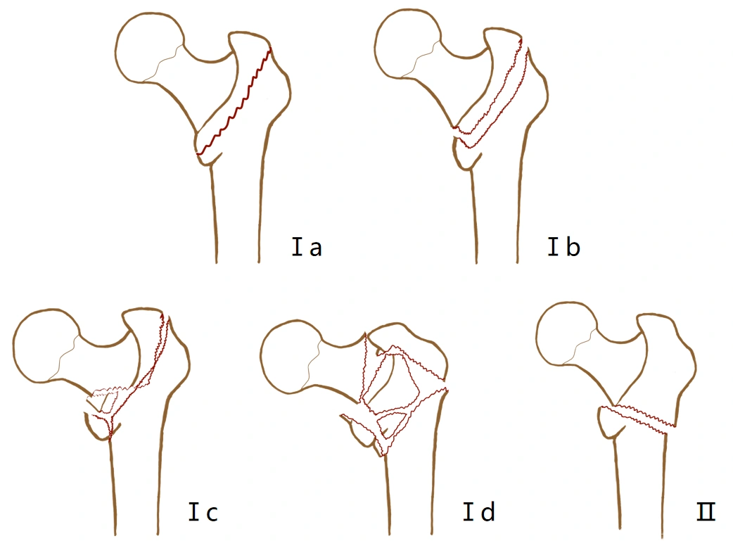
The Evans classification is a traditional system for evaluating intertrochanteric fracture stability. It broadly distinguishes conventional intertrochanteric fracture patterns from reverse-obliquity patterns.
The original classification described Type I fractures with several subtypes based on displacement, fragment configuration, and stability after reduction, and Type II fractures characterized by reverse-obliquity geometry.
Evans Type I: Conventional Intertrochanteric Fractures
Type Ia: A nondisplaced two-part fracture.
Type Ib: A displaced two-part fracture in which medial cortical support remains relatively intact and stability can be restored after reduction.
Type Ic: A fracture with a separate lesser trochanteric fragment or an isolated greater trochanteric fragment, often associated with displacement or varus deformity.
Type Id: A more complex pattern in which both the greater and lesser trochanters become separate fragments, with possible varus displacement.
Types Ia and Ib are generally described as stable patterns in the source classification, while Types Ic and Id are considered unstable.
Evans Type II: Reverse-Obliquity Fractures
In a reverse-obliquity pattern, the fracture line extends from below the greater trochanter toward the medial proximal femur above the lesser trochanter. This geometry may compromise the stability of conventional sliding fixation and can influence implant selection.
The precise terminology and subtype definitions may vary among publications and modified versions of the Evans classification. The original classification reference should be consulted when reproducing a classification diagram.
The Boyd and Griffin classification covers fractures extending from the extracapsular region of the femoral neck to approximately 5 cm below the lesser trochanter.
It describes fracture morphology, comminution, displacement, and the difficulty of achieving and maintaining reduction.
Type I: Simple Intertrochanteric Fractures
These fractures follow the intertrochanteric region from the greater to the lesser trochanter. They are generally relatively simple, with limited comminution and little displacement. Reduction is usually straightforward, and stable fixation may be easier to achieve.
The original source reports this group as approximately 21% of fractures in its series.
Type II: Comminuted Intertrochanteric Fractures
These fractures contain multiple fragments, with the principal fracture involving the intertrochanteric region and multiple areas of cortical disruption. Displacement and comminution can make reduction more difficult, although a stable reduction may be achievable.
Some patterns may appear relatively simple on an anteroposterior radiograph but reveal additional fracture lines on another projection. This illustrates why assessment in more than one plane is important.
The original source reports this group as approximately 36% of fractures in its series.
Type III: Fractures with Subtrochanteric Extension
These fractures involve the proximal femoral shaft at or below the level of the lesser trochanter and may include a large posteromedial comminuted region.
They are often mechanically unstable and can be more difficult to reduce and fix. Treatment may involve greater technical challenges and a higher risk of complications, depending on the fracture pattern and patient characteristics.
The original source reports this group as approximately 28% of fractures in its series.
Type IV: Multiplanar Fractures
These fractures involve the trochanteric region and proximal femoral shaft in at least two planes. The shaft component may be spiral, oblique, or butterfly-shaped.
The complex three-dimensional configuration can complicate reduction and fixation. The surgical plan must address the different fracture planes and provide sufficient stability for healing.
The original source reports this group as approximately 15% of fractures in its series.
These percentages describe the source series and should not be interpreted as universal prevalence estimates. The Boyd and Griffin classification is relatively straightforward but is less commonly used in some contemporary clinical settings than newer classification approaches.
The AO/OTA classification provides a standardized framework for describing proximal femoral fracture morphology and supports communication, research, and treatment planning.
In the traditional AO classification of trochanteric fractures, the major groups include A1, A2, and A3.
AO/OTA 31-A1: Simple Pertrochanteric Fractures
A1 fractures are generally simple two-part pertrochanteric fractures. The medial cortex provides relatively good support, and the lateral cortex remains intact.
Traditional subgroups describe fracture lines that:
Follow the intertrochanteric line
Pass through the greater trochanter
Pass through the lesser trochanter
These patterns are often considered relatively stable when reduction restores adequate cortical support.
AO/OTA 31-A2: Multifragmentary Pertrochanteric Fractures
A2 fractures are multifragmentary. The medial cortex is disrupted at more than one level, while the lateral wall is intact in the traditional classification description.
Subgroups include patterns with one medial fragment, several medial fragments, or fracture extension below the lesser trochanter.
The extent of comminution and the integrity of the lateral wall influence the stability of the fracture and the choice of fixation.
AO/OTA 31-A3: Reverse-Obliquity and Transverse Fractures
A3 fractures include reverse-obliquity, transverse, and more complex patterns involving the lateral cortex.
These fractures may be mechanically unstable and are often considered for intramedullary fixation, depending on the exact fracture geometry and clinical circumstances.
In the traditional classification described in the original article, A1.1 through A2.1 are generally categorized as stable, while A2.2 through A3.3 are categorized as unstable. Actual stability should nevertheless be assessed from the fracture morphology and reduction rather than inferred solely from a subgroup label.
AO/OTA classification is useful for standardized fracture description, statistical analysis, clinical research, and communication among surgeons.
It can also inform implant selection by identifying fracture configurations associated with loss of medial or lateral cortical support.
However, a classification label alone cannot fully predict postoperative stability or outcome. Reduction quality, bone quality, implant design, implant position, and the integrity of the lateral wall must also be considered.
Patients commonly present with pain in the hip or groin following a fall or other traumatic event.
Displaced fractures often prevent the patient from standing or walking, and hip movement may be severely limited by pain. Some patients with nondisplaced fractures may retain limited walking ability despite discomfort.
Because some patients report only thigh or hip pain and may not recall a clear traumatic event, clinicians should consider a possible hip fracture when symptoms and examination findings are suspicious.
The trochanteric region contains vascular cancellous bone. Bleeding around the fracture may produce local swelling and extensive subcutaneous bruising.
Bruising may become more apparent in dependent areas as blood tracks through the soft tissues. The degree of visible bruising does not, by itself, reliably establish the extent or stability of the fracture.
A displaced intertrochanteric fracture may cause shortening and external rotation of the affected lower limb. The degree of deformity generally depends on fracture displacement and the forces acting on the fragments.
Nondisplaced fractures may not produce obvious deformity. The absence of shortening or external rotation therefore does not exclude a fracture.
Assessment should include the history of injury, pain location, swelling, bruising, tenderness, functional impairment, and limb alignment.
Distal neurovascular status should also be assessed. Clinicians should avoid repeatedly provoking pain or attempting unsupported weight bearing when a fracture is suspected.
Compared with many femoral neck fractures, displaced intertrochanteric fractures may produce more obvious external rotation, shortening, and extensive local hematoma. These findings are not sufficiently specific to distinguish the two injuries without imaging.
Plain radiography is the first-line imaging method for suspected intertrochanteric fractures. Assessment commonly includes an anteroposterior pelvic view and appropriate views of the affected hip.
Imaging should be reviewed for:
The direction and extent of the fracture line
Displacement and comminution
Greater and lesser trochanteric fragments
Medial and lateral cortical integrity
Reverse-obliquity or subtrochanteric extension
Bone quality and associated abnormalities
The original article recommends internal rotation for the anteroposterior view to improve visualization. In practice, positioning should be adapted to the patient's pain and injury, and the affected limb should not be forcibly rotated when a fracture is suspected.
A lateral view can provide additional information about posterior fragments and fracture configuration. CT may be useful for selected complex fractures or when the radiographic anatomy is unclear.
If radiographs are negative but clinical suspicion for a hip fracture remains, MRI is recommended when available and appropriate; CT may be considered when MRI is unavailable or contraindicated, in accordance with local guidelines.
Treatment selection should account for fracture stability, bone quality, pre-injury mobility, medical fitness, and the patient's goals of care.
Surgical fixation is the preferred approach for most displaced intertrochanteric fractures when the patient can appropriately undergo surgery. Nonoperative management remains an option in selected circumstances.
Although surgery is commonly preferred, nonoperative management may be necessary when the risks of anesthesia and surgery are considered unacceptable or when treatment aligns better with the patient's goals of care.
The original article describes two conservative approaches.
Approach 1: Early Mobilization with Symptom Control
The approach attributed to Shaftan emphasizes avoiding prolonged bed rest. After appropriate analgesia, patients may be assisted into a wheelchair and mobilized as their condition permits. Assisted transfers and walking with a suitable aid may be introduced when clinically appropriate.
This approach may be considered in selected patients with substantial medical comorbidities. However, it does not guarantee fracture stability or prevent deformity. Patients and caregivers should understand the potential for malunion, shortening, external rotation, and varus deformity.
Approach 2: Skeletal Traction
Skeletal traction has historically been used to correct shortening, external rotation, and varus displacement while maintaining alignment during healing.
The original source describes tibial skeletal traction using approximately 15% of body weight for 8–12 weeks, with serial radiographs used to monitor fracture position and guide adjustment. Subsequent hip movement and partial weight bearing are introduced according to healing.
This is a description of the original source's protocol, not a universal contemporary recommendation. Prolonged traction can cause significant complications and should not be adopted without specialist assessment. Current treatment decisions must account for the patient's condition, available alternatives, and local clinical guidance.
Complication Prevention During Conservative Management
Patients managed without surgery require careful monitoring for complications associated with immobility, including:
Pneumonia and other pulmonary complications
Pressure injuries, particularly over the sacrum and heels
Achilles tendon shortening or contracture
Venous thromboembolism
Muscle wasting and loss of independence
A structured nursing and rehabilitation plan is essential.
Before surgery, clinicians should review the radiographs carefully and determine whether the fracture is stable or whether stability can be restored after reduction.
The main objective is to achieve an acceptable reduction and sufficiently stable fixation to facilitate mobilization.
The strength and durability of fixation depend on several factors:
Bone quality
Fracture morphology
Reduction quality
Implant design
Implant position and technique
Common fixation options include extramedullary systems, such as the dynamic hip screw (DHS) and dynamic condylar screw (DCS), and intramedullary devices, such as the Gamma nail, PFNA, and INTERTAN.
The following sections describe the three methods emphasized in the original article.
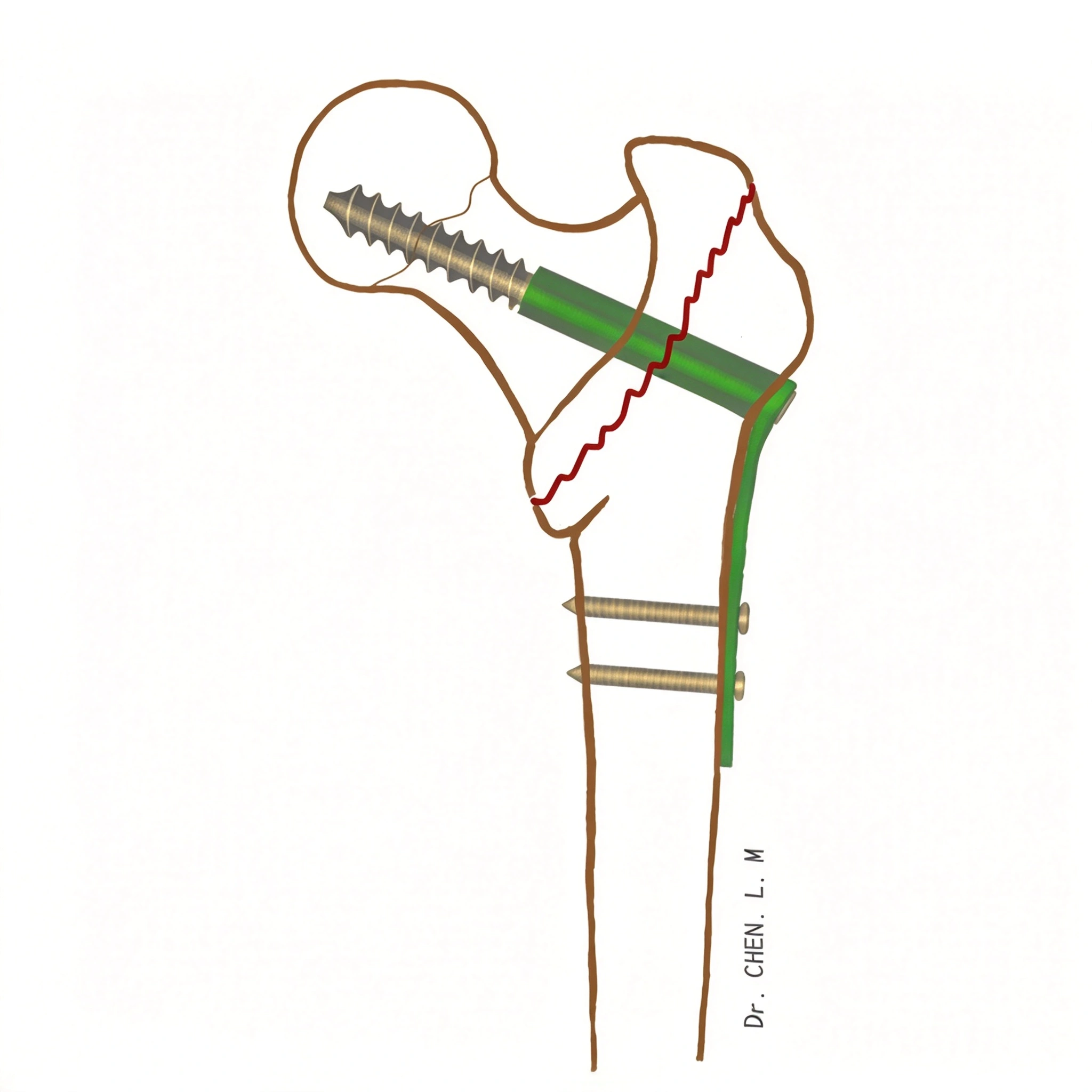
The dynamic hip screw is an extramedullary fixation system that combines a sliding proximal screw with a lateral plate.
General Surgical Procedure
The procedure is commonly performed under spinal or general anesthesia with the patient positioned supine on a fracture table.
Closed fracture reduction is performed under fluoroscopic guidance. Once an acceptable position is obtained, the surgical field is prepared and draped using sterile technique.
A longitudinal incision is made over the lateral aspect of the proximal femur near the greater trochanter. A guidewire is inserted into the femoral head and neck under imaging guidance. The appropriate channel is prepared, and the DHS screw is inserted. The lateral plate is then positioned and secured according to the selected implant system and fracture configuration.
After fluoroscopic confirmation of reduction and implant position, the wound is irrigated and closed in layers. Drain placement depends on the operative findings and the surgeon's preference.
Reduction and Lesser Trochanter Management
DHS fixation is commonly performed after closed reduction. Routine open reduction of an isolated lesser trochanteric fragment is not necessarily required.
Although restoring the posteromedial buttress may improve mechanical support, direct exposure and fixation of the fragment can increase surgical trauma and may disrupt surrounding soft-tissue attachments and blood supply. The decision should depend on fracture morphology and whether satisfactory reduction and stability can be achieved without additional exposure.
Rotational Stability and Additional Screws
The original article discusses whether an additional cannulated screw is required to prevent rotation after DHS fixation. An additional screw should not be considered universally necessary. Its use depends on the fracture pattern, the specific fixation construct, and the surgeon's assessment of rotational stability.
The role of additional fixation should be distinguished from its established use in selected femoral neck or basicervical fracture configurations.
Common Technical Errors
Potential errors include:
Selecting DHS for a fracture pattern poorly suited to conventional sliding fixation, particularly certain reverse-obliquity fractures
Incorrect guidewire or screw position
Inadequate depth or an unfavorable screw trajectory
Loss of fracture reduction during screw insertion
Incorrect positioning or assembly of the screw and side plate
Failure to identify compromised lateral wall support
The proximal screw should be positioned according to accepted surgical principles and the specific implant instructions. A fixed distance from the joint surface should not replace individualized assessment of implant position and fracture morphology.
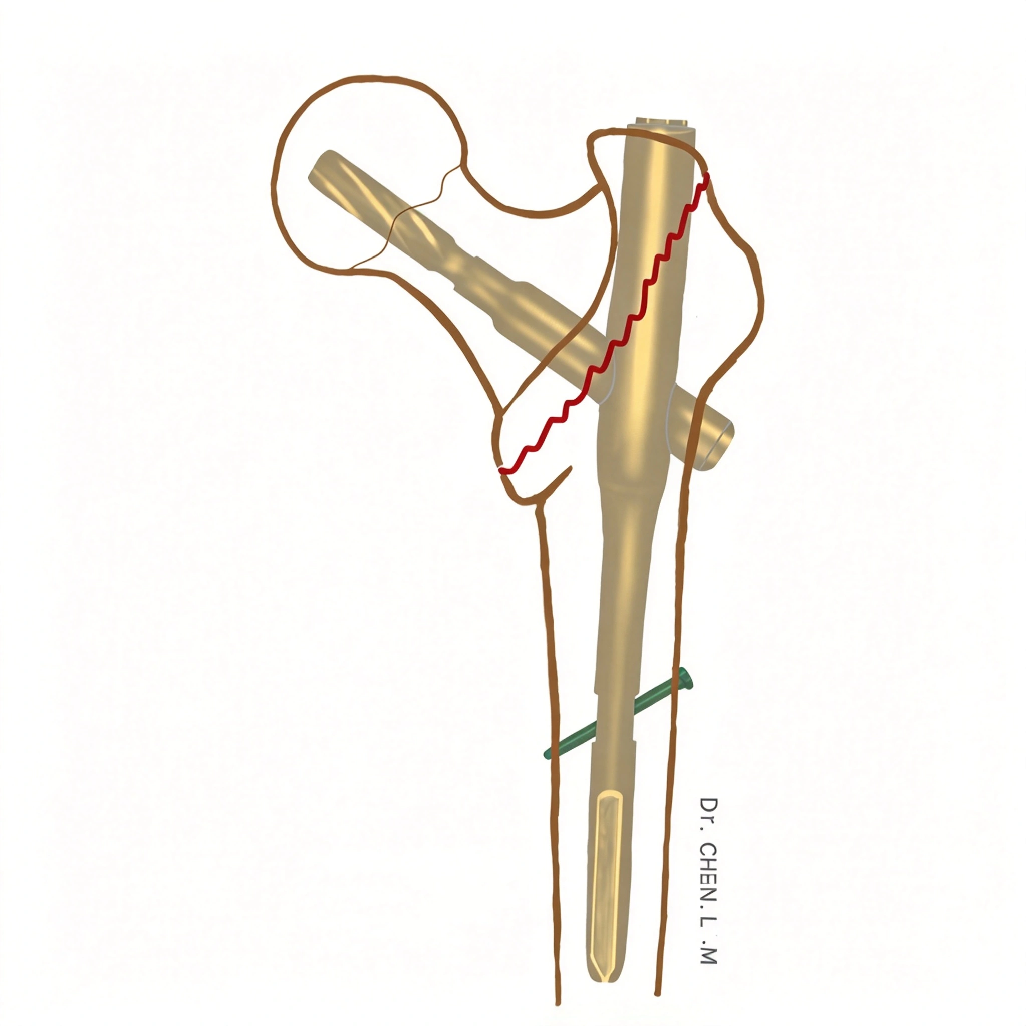
PFNA is an intramedullary fixation system designed for proximal femoral fractures. Many PFNA designs use a helical blade as the proximal fixation element to provide fixation within the femoral head and neck.
General Surgical Procedure
The procedure is commonly performed under spinal or general anesthesia, with the patient supine on a fracture table.
After closed reduction and fluoroscopic confirmation, the proximal femur is approached through a longitudinal incision above the greater trochanter. The entry point is established according to the specific nail design and the patient's anatomy.
The proximal femur is opened, and the main nail is inserted into the femoral canal. Reaming requirements depend on the device and the anatomy; they should follow the manufacturer's instructions rather than a universal rule.
Under fluoroscopic guidance, a guidewire is advanced into the femoral head and neck. Its position is assessed in both anteroposterior and lateral views. The appropriate helical blade is inserted, and the locking mechanism is engaged according to the implant system.
Distal locking is performed using the designated targeting system or an appropriate alternative technique. Final imaging is used to confirm reduction, implant position, and fixation before wound irrigation and closure.
Clinical Applications and Potential Advantages
PFNA is commonly considered for unstable intertrochanteric fractures, including selected patterns represented by Evans Types Ic, Id, and II.
Potential advantages include:
An intramedullary load-sharing configuration
Proximal fixation designed to resist rotation
Applicability to many unstable fracture patterns
A relatively short mechanical lever arm compared with many extramedullary constructs
The original article describes PFNA as offering favorable antirotation performance, potentially shorter operative time, reduced blood loss, and a lower risk of proximal fixation failure in selected settings. These outcomes are not guaranteed and depend on fracture morphology, reduction quality, implant position, bone quality, surgical technique, and the comparator device.
A specific smaller PFNA design may also have different characteristics from other PFNA models. Claims regarding reduced pain or fewer complications should be supported by the relevant clinical evidence before being used as general product claims.
INTERTAN is an intramedullary fixation system developed for proximal femoral fractures, including intertrochanteric fractures. Its distinctive integrated dual-screw configuration is designed to provide proximal fixation and help control rotation and fracture collapse.
General Surgical Procedure
The procedure is commonly performed under spinal or general anesthesia with the patient supine on a fracture table.
Following closed reduction and fluoroscopic confirmation, a longitudinal incision is made above the greater trochanter. The proximal femoral entry point is established in accordance with the specific INTERTAN implant design and surgical technique.
The proximal femur is opened, and the main nail is inserted into the femoral canal. Fluoroscopy is used to confirm nail position and alignment.
A guidewire is then placed into the femoral head and neck. Its position is assessed in the anteroposterior and lateral views, with the intended trajectory and depth confirmed before insertion of the proximal fixation components.
The integrated proximal screw components are inserted according to the manufacturer's technique. Distal locking is then performed using the designated instrumentation or an appropriate alternative method. Final fluoroscopic images are obtained to assess reduction and implant position before wound irrigation and closure.
The exact insertion sequence, screw dimensions, and target measurements should follow the instructions for the specific implant system.
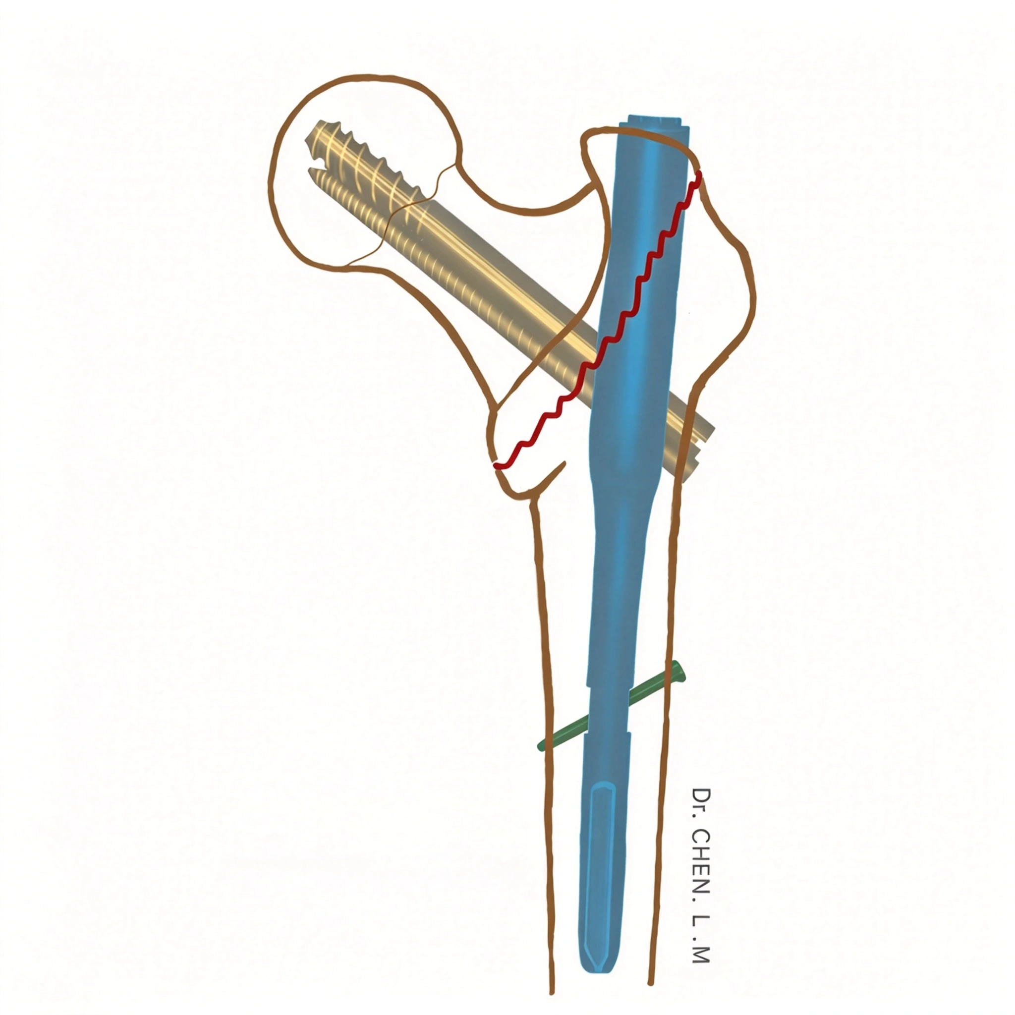
Design Characteristics and Potential Advantages
INTERTAN has a distinctive integrated proximal fixation design. Its nail cross-section and proximal geometry are intended to support insertion and fixation while preserving the surrounding proximal femoral bone to the extent permitted by the anatomy and surgical technique.
The integrated dual-screw configuration is designed to improve rotational control and reduce certain modes of relative screw movement. The original article highlights the potential to reduce the Z-effect observed with some other dual-element proximal femoral nail designs.
INTERTAN may be considered for selected intertrochanteric fracture patterns, including cases in which stable proximal fixation is important in the presence of poor bone quality.
However, no implant design eliminates the risk of fixation failure. Outcomes depend on reduction, implant positioning, bone quality, fracture stability, and surgical technique. Claims of superior outcomes or suitability for all patients with severe osteoporosis require support from appropriate comparative clinical evidence.
DHS, PFNA, and INTERTAN differ in their fixation principles and mechanical configurations. The choice should be based on the fracture pattern and the patient's clinical circumstances rather than the implant name alone.
| Feature | DHS | PFNA | INTERTAN |
|---|---|---|---|
| Fixation configuration | Extramedullary sliding screw and lateral plate | Intramedullary nail with a proximal helical blade in common designs | Intramedullary nail with an integrated proximal dual-screw configuration |
| Proximal fixation principle | Sliding fixation within the femoral head and neck | Proximal fixation designed to provide purchase and rotational control | Integrated dual-screw fixation designed to provide purchase and rotational control |
| Fracture selection | Often suitable for selected stable fracture patterns | Commonly considered for many unstable patterns | Commonly considered for selected intertrochanteric patterns, including some unstable fractures |
| Lateral wall considerations | Integrity of the lateral wall is particularly important | Intramedullary positioning may be advantageous in selected unstable patterns | Intramedullary positioning may be advantageous in selected unstable patterns |
| Main technical considerations | Reduction, screw position, plate placement, and lateral wall support | Reduction, entry point, blade position, and locking | Reduction, entry point, proximal screw positioning, and locking |
| Important limitations | May be unsuitable for certain reverse-obliquity or unstable configurations | Fixation failure remains possible with poor reduction or suboptimal implant position | Fixation failure remains possible despite the integrated screw design |
Clinical interpretation
DHS remains an established option for selected intertrochanteric fractures, particularly when stable reduction and appropriate cortical support can be achieved.
PFNA and INTERTAN are intramedullary alternatives that may be considered for unstable fracture configurations. Their proximal fixation designs differ, and neither should be assumed to be universally superior to the other.
When comparing these devices, clinicians should consider fracture stability, reduction quality, bone quality, implant positioning, operative risks, rehabilitation requirements, and the evidence available for the specific patient group.
Postoperative management aims to restore mobility, prevent medical complications, and support fracture healing.
The original article recommends beginning walking training on the first postoperative day, using a walking aid and partial weight bearing where appropriate. It also describes a plan in which weight bearing during the first week reaches approximately 50% of the load permitted by the normal limb.
These figures represent the original source's rehabilitation approach, not a universal protocol. The appropriate weight-bearing plan depends on fracture morphology, reduction quality, fixation stability, intraoperative findings, and the treating surgeon's instructions.
Early mobilization should be encouraged when medically and mechanically appropriate. Restrictions that are not supported by the patient's fracture or fixation circumstances may complicate rehabilitation, but unrestricted weight bearing should not be prescribed automatically for every case.
Patients with weak upper limbs or concomitant upper-extremity fractures may find it difficult to use a walker or crutches. Rehabilitation aids and assistance should be selected according to individual functional limitations.
Loading of the hip does not occur only during walking. Transfers, repositioning, sitting, and other everyday activities also generate forces around the hip.
Muscle contraction during bed exercises can likewise load the proximal femur. Consequently, prolonged bed rest does not eliminate mechanical loading and may increase the risks associated with immobility.
Clinical studies and guidelines should be reviewed when determining whether unrestricted or restricted weight bearing is appropriate for a particular fracture and fixation construct. The final plan should be individualized rather than based on a blanket rule.
Venous thromboembolism prevention is an important component of postoperative care after hip fracture surgery.
Depending on the patient's medical condition and bleeding risk, preventive measures may include:
Pharmacological prophylaxis, such as low-molecular-weight heparin when appropriate
Mechanical compression devices
Graduated compression stockings when indicated
Early mobilization and appropriate rehabilitation
The choice, timing, and duration of prophylaxis should follow current guidelines and individual risk assessment.
Postoperative follow-up should assess pain, wound healing, functional recovery, fracture alignment, and evidence of healing. Imaging may be required according to the clinical course and the surgeon's assessment.
Patients should receive clear instructions on safe transfers, use of walking aids, prescribed weight-bearing limits, and signs that require prompt medical review.
The original article describes traditional Chinese medicine (TCM) approaches organized according to the stages of fracture recovery. These approaches should be understood as traditional therapeutic frameworks rather than established substitutes for fracture fixation, rehabilitation, or evidence-based medical management.
The traditional framework describes the early stage in terms of injury to the muscles, bones, and meridians, with an emphasis on blood stasis, swelling, and pain.
Traditional prescriptions mentioned in the source include modified Heying Zhitong Tang and Fuyuan Huoxue Tang. Other formulas or herbs may be added in traditional practice according to the patient's presentation.
The clinical use of these prescriptions should be assessed individually. Evidence for effectiveness and safety in patients with hip fractures varies, and potential interactions with anticoagulants, anesthetic drugs, and other medications must be considered.
The source describes the middle stage as a period in which swelling and pain may gradually improve while bone healing remains incomplete.
Traditional treatment principles emphasize supporting bone and tendon recovery. Formulas mentioned include modified Xin Shang Xu Duan Tang and Taohong Siwu Tang.
These treatments should not replace appropriate postoperative follow-up, rehabilitation, nutritional support, or assessment for complications.
The later stage is traditionally associated with callus formation and progressive fracture healing. The source emphasizes strengthening muscles and bones and supporting the body's recovery.
Traditional formulas mentioned include Liuwei Dihuang Tang, Bazhen Tang, and Huqian Wan.
The use of herbal preparations requires appropriate professional assessment, especially in older adults with multiple comorbidities or those taking several medications. Fracture healing should be assessed clinically and radiographically rather than inferred solely from the elapsed time or traditional treatment stage.
Osteoporotic intertrochanteric fractures are closely associated with reduced bone strength and falls. Prevention should address both factors.
Important strategies include:
Assessment and treatment of osteoporosis
Adequate dietary protein, calcium, and vitamin D when indicated
Regular physical activity appropriate to the patient's abilities
Strength and balance training
Review of medications that may increase fall risk
Assessment of vision, gait, and environmental hazards
Home modifications to reduce the risk of falls
Follow-up to assess bone health and future fracture risk
For patients who have already sustained a hip fracture, secondary fracture prevention is an important part of long-term care.
Intertrochanteric femur fractures require careful evaluation of fracture morphology, mechanical stability, bone quality, and the patient's overall clinical condition. The Evans, Boyd and Griffin, and AO/OTA classification systems help describe fracture patterns and inform treatment planning, although no classification system can fully replace individualized assessment.
For patients undergoing surgery, the main objective is to restore acceptable alignment and achieve stable fixation that supports appropriate mobilization. DHS, PFNA, and INTERTAN are important fixation options with different mechanical configurations and indications. DHS may be appropriate for selected stable patterns, while intramedullary systems may be considered for many unstable fractures. The final choice depends on fracture characteristics, implant design, reduction quality, bone quality, and clinical evidence.
Postoperative management should emphasize early mobilization when appropriate, individualized rehabilitation, prevention of venous thromboembolism and other complications, and assessment for osteoporosis. Traditional Chinese medicine approaches may be discussed as adjunctive care where appropriate, but their safety and evidence should be evaluated and they should not replace established fracture treatment.
A comprehensive, patient-centered approach can help support fracture healing, reduce complications, preserve mobility, and improve quality of life.
National Institute for Health and Care Excellence (NICE). Hip fracture: management (CG124).
https://www.nice.org.uk/guidance/cg124/chapter/recommendations
American Academy of Orthopaedic Surgeons (AAOS). Management of Hip Fractures in Older Adults: Clinical Practice Guideline.
https://new.aaos.org/quality/quality-programs/hip-fractures-in-older-adults/
AO Foundation. AO/OTA Fracture and Dislocation Classification.
https://www.aofoundation.org/trauma/clinical-library-and-tools/journals-and-publications
AO Surgery Reference. Proximal femur fractures: surgical approaches and fixation principles.
https://surgeryreference.aofoundation.org/orthopedic-trauma/adult-trauma/proximal-femur
National Library of Medicine. Orthogeriatrics and the management of hip fractures in older adults.
https://www.ncbi.nlm.nih.gov/books/NBK565572/
Medical Disclaimer: This article is intended for educational and professional information purposes only. It does not replace individualized clinical assessment, the treating surgeon's judgment, applicable clinical guidelines, or the instructions for use supplied with a specific orthopaedic implant. Surgical procedures and postoperative weight-bearing protocols must be determined by qualified healthcare professionals.


