English


Views: 0 Author: Site Editor Publish Time: 2026-10-16 Origin: Site
Introduction
The scapholunate interosseous ligament (SLIL) is a major stabilizer of the wrist, connecting the scaphoid and lunate bones within the proximal carpal row. Injury to this ligament can disrupt normal carpal alignment, producing wrist pain, mechanical clicking, reduced grip endurance, and progressive instability.
Early recognition is important because selected acute injuries remain suitable for direct ligament repair. When the dorsal portion of the SLIL is completely torn but the tissue remains repairable, surgical treatment may involve open reduction, direct ligament reattachment, and temporary K-wire fixation. Additional procedures, such as dorsal capsulodesis, may be considered when indicated by the condition of the surrounding stabilizers.
Chronic injuries present a different challenge. Ligament tissue may become attenuated or irreparable, the scapholunate interval may remain widened, and dorsal intercalated segment instability (DISI) or degenerative changes may develop. In these situations, reconstruction or other procedures may be more appropriate than primary repair.
This article reviews SLIL anatomy, clinical presentation, diagnostic examination, and the principles of open repair for selected acute injuries, with an emphasis on restoring carpal alignment while protecting the wrist’s remaining stabilizing structures.
The SLIL connects the scaphoid and lunate and is commonly described as having three regions:
Dorsal portion: A thick, strong bundle of collagen fibers that provides major resistance to scapholunate separation and abnormal translation or rotation.
Volar portion: An obliquely oriented fibrous component that contributes to rotational control and coordination between the scaphoid and lunate.
Proximal membranous portion: A relatively thin fibrocartilaginous region that separates the radiocarpal joint from the midcarpal joint.
Although these components form a continuous ligament complex, they do not contribute equally to mechanical stability. The dorsal portion is generally considered the strongest and a particularly important restraint against scapholunate instability. The volar portion contributes to rotational control, while the proximal membranous portion has less structural strength.
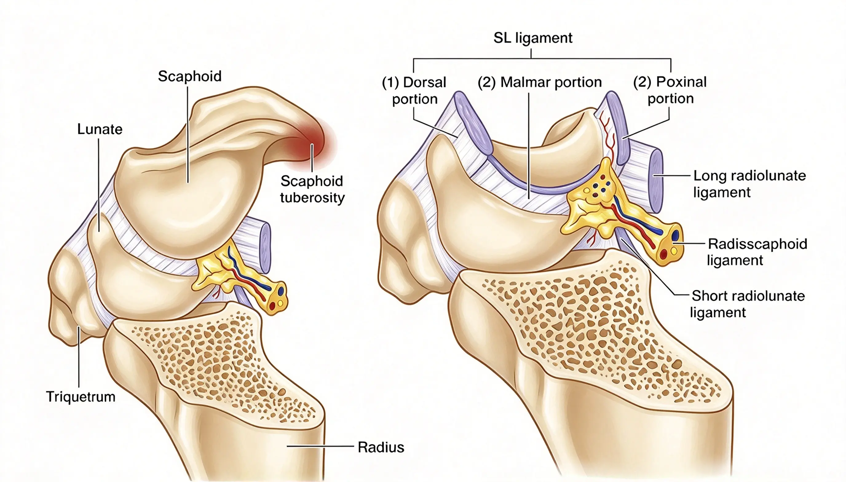
The SLIL does not stabilize the wrist in isolation. Extrinsic and other intrinsic ligament structures contribute to normal carpal motion and help maintain the relationship between the scaphoid and lunate.
Relevant structures include the dorsal intercarpal ligament, dorsal radiocarpal ligament, radioscaphocapitate ligament, and scaphotrapeziotrapezoid ligament complex.
When the SLIL is disrupted, the remaining stabilizers may partially compensate for the injury. If these structures are also compromised, scapholunate instability can become more pronounced or progress from dynamic to static malalignment.
This distinction is important when selecting a treatment. A repairable dorsal SLIL tear with preserved secondary stabilizers is different from a chronic injury involving multiple stabilizing structures.
The scaphoid and lunate normally move in a coordinated manner during wrist motion. The scaphoid tends to flex, while the lunate’s motion is influenced by the surrounding ligament network and adjacent carpal bones.
When the SLIL loses its restraining function, the scaphoid may flex and rotate abnormally, while the lunate may extend. Depending on the integrity of the secondary stabilizers and the severity of the injury, this can produce scapholunate widening and a DISI pattern.
Persistent abnormal loading may contribute to progressive cartilage damage and scapholunate advanced collapse (SLAC) wrist in some patients. However, not every SLIL injury progresses to arthritis, and the natural history varies with injury severity, associated damage, and treatment.
Patients with an SLIL injury may report:
Pain over the dorsal or dorsoradial wrist, particularly during loading or gripping.
Mechanical clicking, clunking, or a sensation of wrist shifting.
Reduced grip endurance, especially after repeated gripping.
Localized swelling or tenderness over the scapholunate interval.
Difficulty performing activities that require wrist extension under load.
Wrist range of motion and initial grip strength may remain relatively well preserved, particularly in less severe injuries. Consequently, a patient can have a clinically significant ligament injury despite a relatively normal initial assessment.
An injury history involving a fall onto an extended wrist or another loading event should raise suspicion when pain is localized to the scapholunate interval.
Examination should include inspection for swelling, palpation of the scapholunate interval, assessment of wrist motion, and evaluation of grip strength and carpal stability.
Tenderness is often found over the dorsal scapholunate interval, near the region just distal to Lister’s tubercle. Symptoms may increase during wrist movement or repetitive gripping.
A structured examination should also consider associated injuries, including occult fractures, other intrinsic carpal ligament injuries, and injuries involving the triangular fibrocartilage complex when the clinical presentation suggests them.
The Watson test, also known as the scaphoid shift test, is a provocative maneuver used to assess scaphoid instability.
The examiner stabilizes the wrist and applies pressure over the palmar prominence of the scaphoid while moving the wrist from ulnar deviation and slight extension toward radial deviation and slight flexion. If the scaphoid is unstable, it may subluxate dorsally. Releasing the pressure can allow it to return toward its normal position, sometimes producing a painful clunk.
A painful, reproducible clunk can support the suspicion of scapholunate instability, but the test is not diagnostic on its own. Some individuals without a clinically significant SLIL tear may demonstrate a positive maneuver, and a negative test does not exclude an injury. One arthroscopy-correlated study found limited sensitivity and specificity, reinforcing the need to interpret the test alongside imaging and the overall clinical assessment.
Initial imaging generally includes standard wrist radiographs. Depending on the injury, additional views or stress radiographs may help identify dynamic widening of the scapholunate interval.
In dynamic instability, the interval may appear relatively normal on routine neutral images but widen under stress. In static instability, widening or abnormal carpal alignment may already be visible on standard radiographs.
The scapholunate gap, scapholunate angle, carpal alignment, and any evidence of degenerative change should be assessed together rather than relying on a single measurement.
MRI or MR arthrography may help evaluate ligament injury and associated soft-tissue abnormalities. However, imaging findings must be correlated with symptoms and physical examination because some tears or degenerative changes may be difficult to interpret in isolation.
Wrist arthroscopy can directly assess the SLIL, the extent of instability, and the condition of the articular surfaces. It may be particularly valuable when noninvasive studies do not adequately characterize the injury or when operative treatment is being considered.
Treatment planning should account for several factors:
Time since injury: Acute injuries may retain tissue suitable for direct repair, while chronic injuries may require reconstruction.
Ligament quality: The remaining tissue must be capable of holding a repair.
Reducibility: The scaphoid and lunate should be assessed for whether normal or acceptable alignment can be restored.
Secondary stabilizers: Associated ligament damage can influence the stability of the repair.
Cartilage condition: Established degenerative changes may change the range of appropriate surgical options.
The commonly cited acute treatment window is short, often measured in the first few weeks after injury. However, the exact timing is not an absolute rule; intraoperative tissue quality, reducibility, and cartilage status remain important.
Open direct repair may be considered when the SLIL is acutely disrupted, the dorsal component remains repairable, the scapholunate relationship can be reduced, and there is no advanced cartilage damage.
Many injuries involve avulsion of the ligament from the scaphoid, sometimes with a small attached bone fragment. Depending on the tear pattern, the ligament may be reattached with a suture anchor or transosseous sutures. Temporary K-wire fixation can protect the repair while it heals.
The principal goals are to:
Restore acceptable scaphoid–lunate alignment.
Reattach repairable ligament tissue.
Protect the repair during early healing.
Preserve the surrounding capsuloligamentous structures.
Reduce the risk of persistent instability and abnormal carpal mechanics.
Direct repair primarily addresses repairable tissue. It should not be assumed to restore every damaged component of the scapholunate stabilizing system.
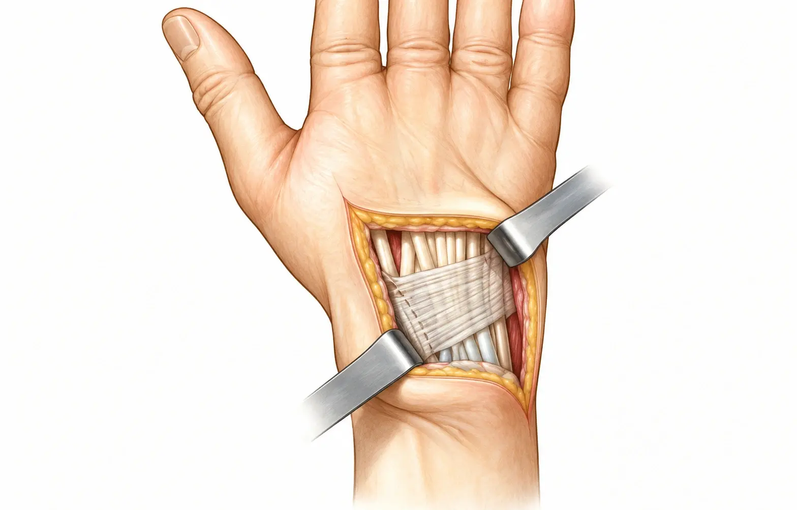
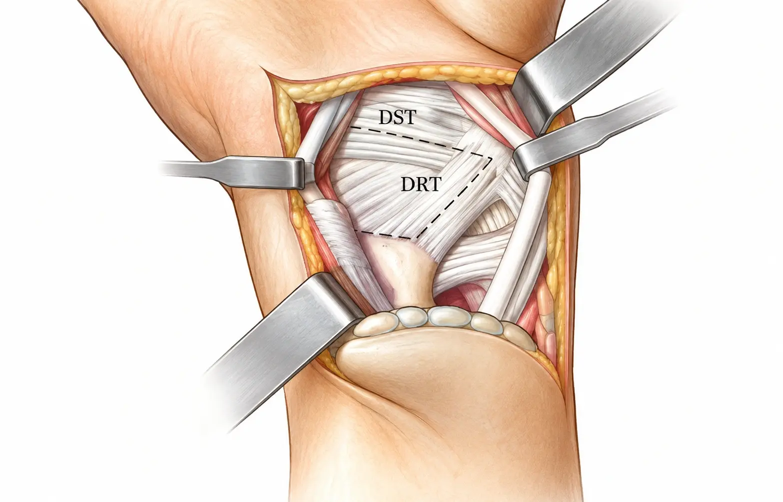
A dorsal wrist approach is commonly used to access the dorsal SLIL. The incision and capsular exposure should provide adequate visualization while preserving important ligamentous and extensor structures whenever possible.
A ligament-sparing dorsal capsulotomy may be used according to the selected technique. Exposure typically involves careful handling of the extensor tendons and the dorsal capsule.
The precise approach varies with the surgeon’s preferred method, the injury pattern, and whether associated pathology requires additional assessment.
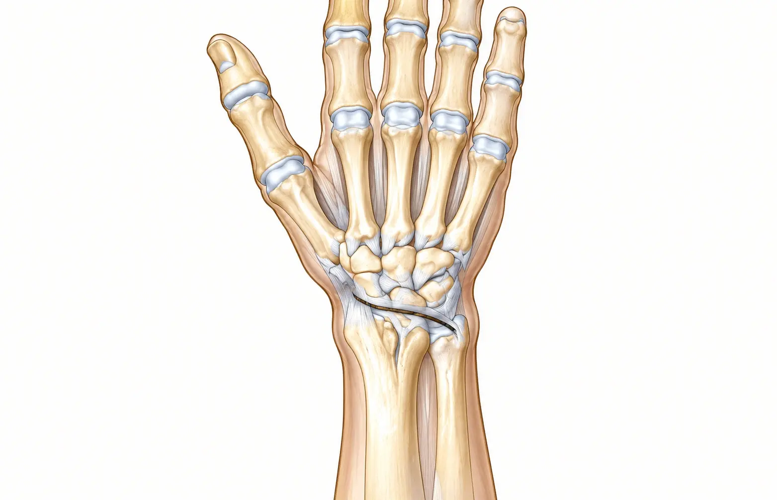
▲ Dorsal curvilinear or longitudinal wrist incision
After exposure, the surgeon identifies the disrupted ligament and evaluates its attachment site and tissue quality. If the ligament is avulsed from the scaphoid or lunate, the presence and size of any attached bone fragment should be assessed.
For selected avulsion injuries, the receiving bone surface may be prepared to support reattachment. The original described technique includes preparing a small bony recess at the scaphoid attachment region and creating bone tunnels for transosseous sutures.
These steps are technique-specific. The extent of bone preparation should be appropriate to the tear pattern and should avoid unnecessary damage to the articular surface or remaining ligament attachments.
When the dorsal SLIL is repairable, it may be reattached using suture anchors or transosseous sutures. A mattress-type suture configuration is one option for capturing the ligament tissue, depending on its quality and the chosen technique.
The aim is to restore the ligament attachment while maintaining the reduced relationship between the scaphoid and lunate. A small avulsion fragment may be repaired directly when its size, location, and bone quality permit secure fixation.
No single fixation method is appropriate for every tear. The choice between anchors, transosseous fixation, and fragment fixation depends on the available tissue, bone anatomy, and the surgeon’s assessment.
After the ligament has been repaired, the scaphoid and lunate must be held in an acceptable relationship while the repair heals.
Temporary K-wires may be used to protect the repair and maintain reduction. Additional fixation across the scaphocapitate joint may be considered according to the degree of instability and the operative plan.
If residual DISI alignment is present, the surgeon must reassess the reduction and the position of the lunate before completing fixation. The appropriate correction depends on the underlying instability and cannot be determined by one maneuver alone.
K-wire placement requires careful attention to surrounding structures, including tendons, articular surfaces, and nearby sensory nerves. Fluoroscopic or other appropriate intraoperative imaging can help verify alignment and fixation.
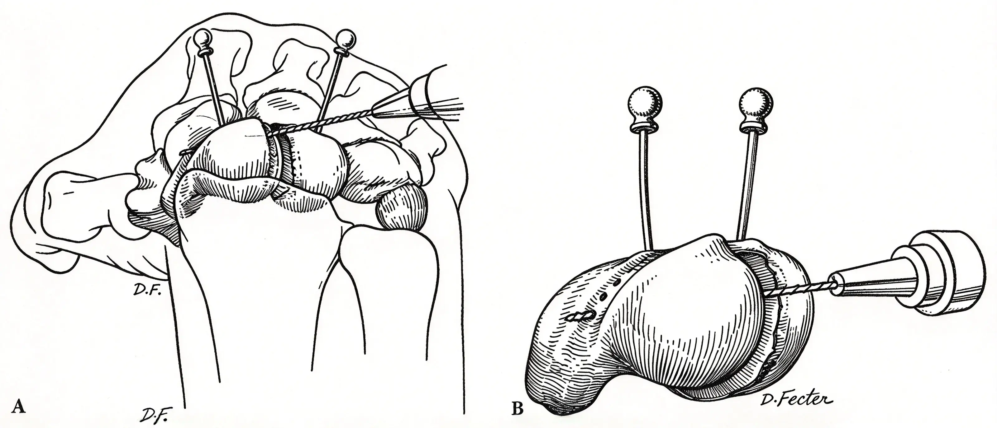
In selected cases, the dorsal capsule and associated extrinsic ligaments may provide additional support after direct SLIL repair. Dorsal capsulodesis uses a portion of the dorsal capsuloligamentous tissue to help limit abnormal scaphoid motion or support carpal alignment.
One described technique uses a proximally based dorsal capsular flap that is mobilized and secured to the scaphoid region. Other methods use portions of the dorsal intercarpal ligament or related tissues, depending on the specific procedure.
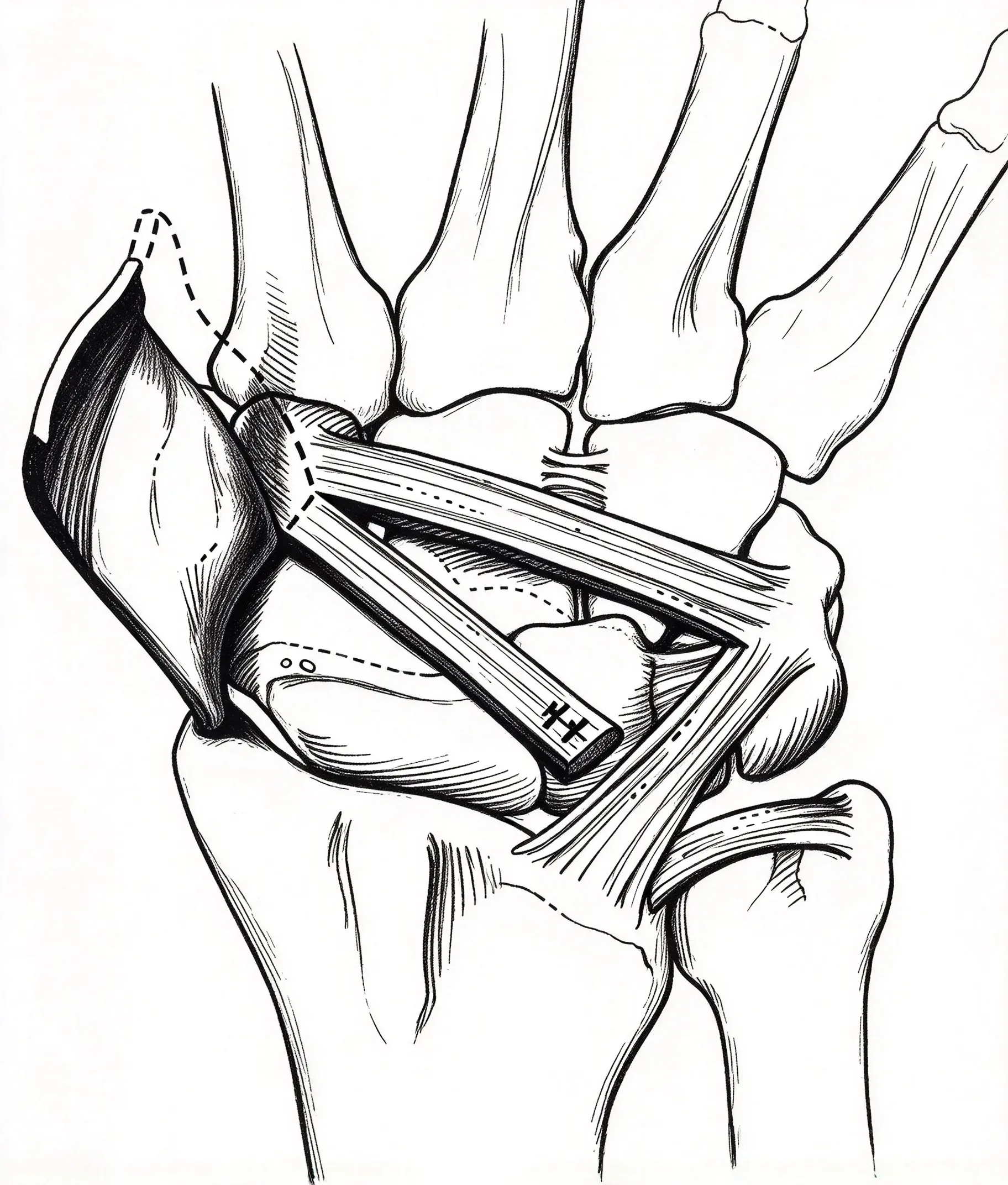
Capsulodesis may be considered when the dorsal capsule remains continuous but lax, or when the surgeon judges that additional soft-tissue support is appropriate after reduction and repair.
It is not a substitute for repairing an otherwise repairable SLIL when direct repair is indicated. Nor does it guarantee long-term maintenance of alignment. Published reviews describe variable outcomes, including potential loss of wrist motion and persistent or recurrent instability. The decision should therefore be individualized.
Following direct SLIL repair with temporary K-wire fixation, wrist immobilization is generally required while the repair heals. The duration depends on the fixation construct, tissue quality, associated procedures, and the treating surgeon’s protocol.
Finger motion and appropriate hand exercises are often initiated early to reduce stiffness, provided they do not jeopardize the repair.
Wrist range-of-motion exercises and strengthening are introduced progressively after the surgeon determines that the repair is sufficiently protected. Loading, forceful gripping, and weight-bearing through the hand should be restricted until adequate healing and stability have been confirmed.
Follow-up assessment may include clinical examination and radiographs to evaluate alignment, fixation, and the progression of healing. K-wire removal timing and rehabilitation progression should follow the specific operative plan rather than a universal schedule.
Possible complications include persistent or recurrent scapholunate instability, stiffness, loss of grip strength, pin-related problems, infection, and progression of degenerative changes. Some patients may continue to experience pain despite restoration of alignment.
Patients should be counseled that ligament repair aims to improve stability and symptoms but cannot guarantee normal wrist mechanics or prevent all future arthritis.
The distinction between acute and chronic injury is central to treatment selection.
| Clinical feature | Acute, repairable injury | Chronic or irreparable injury |
|---|---|---|
| Ligament tissue | May retain sufficient quality for direct repair | May be attenuated, scarred, or irreparable |
| Alignment | Often reducible in selected cases | May involve fixed or recurrent malalignment |
| Common surgical consideration | Direct repair with temporary fixation, with adjunctive stabilization when indicated | Ligament reconstruction, selected capsulodesis procedures, or other procedures based on cartilage and stability |
| Key decision factors | Tissue quality, reducibility, secondary stabilizers, cartilage status | Reducibility, residual ligament function, secondary stabilizers, cartilage condition, symptoms |
Chronic injuries are not managed by a single standard procedure. Depending on the degree of instability and the condition of the articular surfaces, options may include ligament reconstruction, selected capsulodesis techniques, or salvage procedures for advanced degenerative disease.
The scapholunate interosseous ligament connects the scaphoid and lunate bones in the wrist. It helps coordinate carpal motion and maintain stability between these two bones.
Common symptoms include dorsal wrist pain, tenderness over the scapholunate interval, clicking or clunking with movement, and reduced grip endurance. Symptoms and examination findings vary according to injury severity.
The Watson test is a clinical maneuver that assesses scaphoid instability by applying pressure over the scaphoid while moving the wrist. A painful clunk may support the diagnosis, but the test alone cannot reliably confirm or exclude an SLIL injury.
Direct repair may be considered for selected acute injuries in which the dorsal ligament remains repairable, the carpal relationship can be reduced, and there is no advanced cartilage damage. Chronic or irreparable injuries may require reconstruction or another procedure.
Temporary K-wires can protect the repair by maintaining the reduced relationship between the scaphoid and lunate during early healing. The number and configuration of wires depend on the injury and the operative plan.
No. Dorsal capsulodesis is an adjunct considered in selected cases. Its potential benefits must be weighed against limitations such as stiffness, reduced motion, and the possibility of recurrent instability.
Some untreated or persistently unstable injuries progress to degenerative changes, including SLAC wrist. However, progression is not inevitable, and outcomes depend on injury severity, carpal alignment, cartilage condition, and other factors.
Scapholunate ligament injury can compromise wrist stability and produce persistent pain, mechanical symptoms, and abnormal carpal alignment. Accurate diagnosis requires a combination of clinical examination, appropriate imaging, and, when indicated, direct assessment of the ligament and articular surfaces.
For selected acute injuries with repairable dorsal ligament tissue, open reduction and direct SLIL repair with temporary K-wire fixation may restore the scapholunate relationship. Dorsal capsulodesis or other adjunctive stabilization may be considered according to the condition of the surrounding structures.
Chronic or irreparable injuries require a different strategy based on reducibility, secondary stabilizer integrity, and cartilage status. Ultimately, successful management depends on matching the procedure to the injury stage, preserving functional tissue, and protecting the repair through appropriate rehabilitation.
Andersson JK. Treatment of scapholunate ligament injury: Current concepts. EFORT Open Reviews. 2017;2:382–393. Read the article.
Scapholunate ligament injuries: a review of current concepts. Journal of Hand Surgery. Read the review.
Optimal management of scapholunate ligament injuries. Orthopedic Research and Reviews. 2018. Read the article.
Current Role of Open Reconstruction of the Scapholunate Ligament. Journal of Wrist Surgery. 2013;2(2):116–125. Read the article.
Management of chronic scapholunate ligament injury. Read the review.
Relevance of the Scaphoid Shift Test for the Investigation of Scapholunate Ligament Injuries. Read the study.


