
Views: 0 Author: Site Editor Publish Time: 2026-08-14 Origin: Site
“Take a Broden’s view.”
For orthopedic surgeons treating calcaneal fractures, this instruction may sound routine. After fixation of a calcaneal fracture, the C-arm is positioned, the foot is rotated, and a series of oblique radiographs is obtained to assess the posterior facet of the subtalar joint.
But why is this projection called Broden’s view?
Who was Broden, and why has his name remained part of orthopedic imaging for more than seven decades?
The answer goes back to 1949, when Swedish radiologist Bror Broden described a series of specialized oblique radiographic projections designed to improve visualization of the posterior articular surface of the calcaneus.
Although modern computed tomography (CT), intraoperative 3D imaging, navigation, and subtalar arthroscopy have transformed calcaneal fracture management, the fundamental principle behind Broden’s view remains highly relevant:
Radiographic imaging becomes more useful when the projection is designed according to the three-dimensional anatomy of the joint.
This article reviews the history, anatomical principles, projection technique, clinical applications, and modern relevance of Broden’s view in calcaneal fracture surgery.
Before Broden’s work, standard calcaneal radiographic assessment primarily relied on lateral and axial or semi-axial projections.
These views are useful for evaluating:
Calcaneal height
Calcaneal length
Böhler’s angle
Gissane’s angle
Overall fracture displacement
Tuberosity position
Gross subtalar joint abnormalities
However, complex intra-articular calcaneal fractures present a much more difficult imaging problem.
The posterior facet of the subtalar joint is a curved three-dimensional surface. A conventional lateral projection cannot adequately demonstrate the entire articular surface because the X-ray beam does not align optimally with the geometry of the facet.
This becomes particularly important in displaced intra-articular calcaneal fractures, where restoration of the posterior facet is a major objective of surgical reduction.
In the late 1940s, Bror Broden recognized that conventional calcaneal projections provided inadequate information about the posterior articular surface.
His approach was fundamentally anatomical rather than technological.
Instead of simply adding another radiographic angle, Broden first analyzed the three-dimensional geometry of the calcaneus and then designed projections based on that anatomy.
This concept ultimately led to what is now widely known as Broden’s view.
Fig 1.Brodén's conical model of the calcaneal posterior facet: the base faces posterolaterally and the apex anteromedially,
at a 45° angle to the foot's long axis.
The posterior facet of the calcaneus forms an important component of the subtalar joint.
Its curved morphology makes it difficult to visualize completely on a conventional two-dimensional radiograph.
Broden described the posterior articular surface as having a geometry that could be approximated to a portion of a cone.
The broad portion of this geometric model is oriented posterolaterally, while the apex is directed anteromedially.
The central axis of this configuration has an important relationship with the longitudinal axis of the foot.
The key principle is simple:
To obtain a clearer projection of a curved articular surface, the X-ray beam should be positioned as close as possible to perpendicular to the relevant surface.
This explains why a standard lateral radiograph cannot provide complete visualization of the posterior facet.
The projection angle must change.
Broden therefore developed oblique projections in which the foot and X-ray tube are positioned at specific angles to obtain sequential images of different portions of the subtalar joint.
This anatomical reasoning remains one of the most elegant aspects of the original technique.
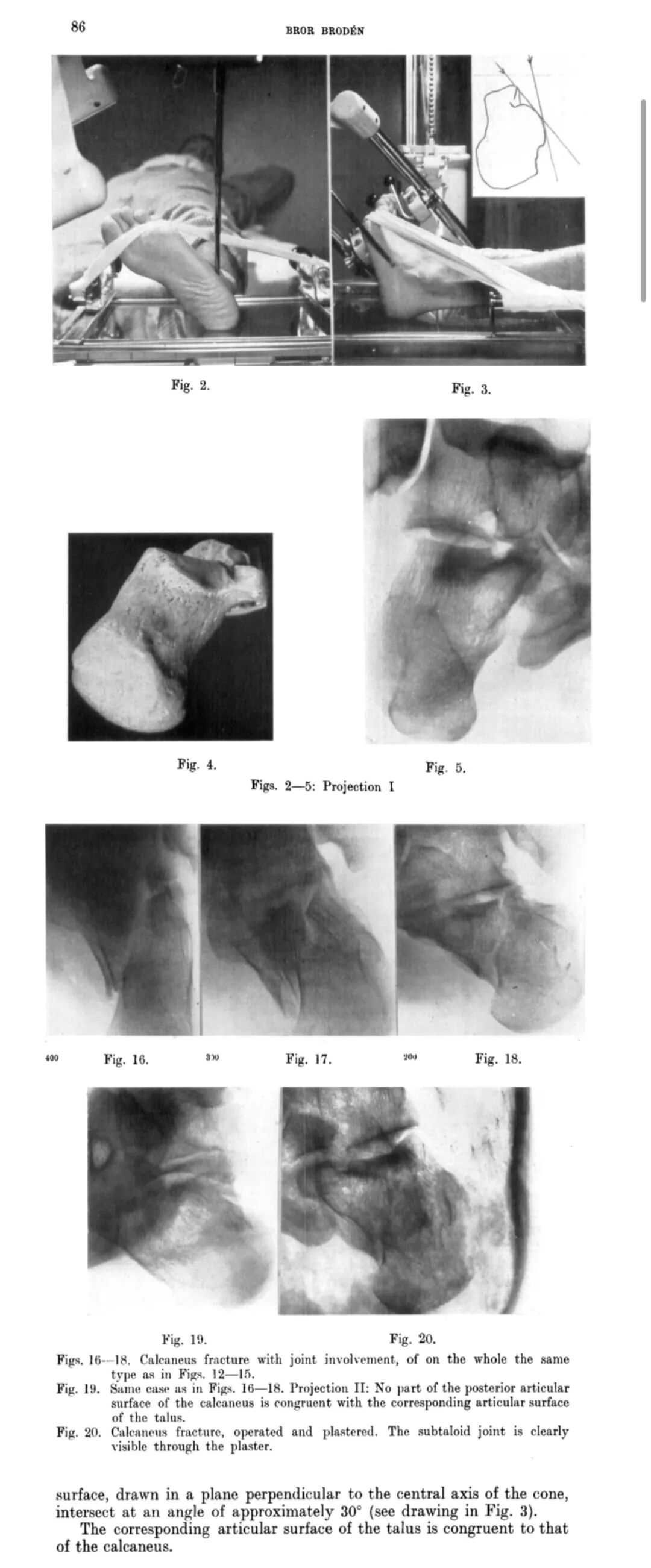
Broden’s view is not necessarily a single radiographic image.
It traditionally refers to a series of oblique radiographic projections designed to visualize the posterior facet of the subtalar joint from different angles.
The technique generally involves:
Supine patient positioning
Approximately 45° internal rotation of the foot
The ankle maintained near a neutral position
The central beam directed toward the region just inferior to the lateral malleolus
Progressive cephalad tube angulation
A commonly described sequence uses tube angulations of approximately:
40°
30°
20°
10°
The different projections demonstrate different portions of the posterior facet.
The posterior facet is curved rather than flat.
Therefore, a single projection cannot necessarily demonstrate its entire surface without overlapping structures.
Changing the tube angle allows the surgeon or radiologist to progressively visualize different regions of the articular surface.
In practical terms:
Higher tube angulation → relatively more anterior visualization
Lower tube angulation → relatively more posterior visualization
The exact appearance depends on patient positioning, fracture morphology, and imaging equipment.
The patient is generally positioned supine.
The affected foot is internally rotated approximately 45° while maintaining the ankle in a comfortable neutral or near-neutral position.
This positioning helps orient the subtalar joint appropriately relative to the X-ray beam.
The C-arm is positioned so that the central beam is directed toward the lateral hindfoot.
The tube is then progressively angled in a cephalad direction.
A commonly used sequence consists of four projections at approximately 40°, 30°, 20°, and 10°.
One practical advantage of the technique is that it does not require aggressive ankle dorsiflexion.
This is particularly useful in patients with:
Acute calcaneal fractures
Severe swelling
Postoperative pain
External fixation
Splints or casts
Minimizing unnecessary manipulation can improve patient comfort and facilitate intraoperative imaging.
Broden’s view is particularly useful for evaluating the posterior facet of the subtalar joint.
It can assist in assessing:
The surgeon can evaluate whether the major articular fragments have been restored to an acceptable relationship.
Residual displacement or step-off of the posterior facet may be visible on appropriately positioned projections.
The relationship between the posterior facet of the calcaneus and the talus can be evaluated.
Broden’s projections can help assess the position of major intra-articular fracture fragments after reduction and fixation.
After insertion of plates and screws, Broden’s view may help confirm that:
The posterior facet remains reduced
Hardware has not significantly displaced the reduction
Screws are appropriately positioned
The subtalar joint remains congruent
Displaced intra-articular calcaneal fractures frequently involve the posterior facet.
Restoring this surface is one of the key objectives of operative treatment.
An inadequate reduction may contribute to:
Post-traumatic subtalar arthritis
Persistent hindfoot pain
Reduced subtalar motion
Altered hindfoot biomechanics
Difficulty with later reconstruction
For this reason, intraoperative assessment of the posterior facet is important.
During open reduction and internal fixation (ORIF), Broden’s view can be incorporated into intraoperative fluoroscopy.
The typical workflow may include:
Fracture reduction
Temporary fixation
Posterior facet assessment
Definitive plate and screw fixation
Repeat Broden projections
Assessment of subtalar joint congruity
Final hardware evaluation
The technique can therefore function as an additional intraoperative quality-control step.
Different radiographic projections provide different information.
| Imaging View | Main Clinical Information |
|---|---|
| Lateral view | Calcaneal height, length, Böhler’s angle, Gissane’s angle |
| Axial/Harris view | Calcaneal width, tuberosity alignment, varus/valgus |
| Broden’s view | Posterior facet and subtalar joint congruity |
| CT | Three-dimensional fracture anatomy and fragment displacement |
No single projection provides a complete assessment of a complex calcaneal fracture.
Instead, these imaging modalities should be considered complementary.
Modern CT provides substantially more anatomical information than conventional radiography.
CT can demonstrate:
Fracture classification
Posterior facet displacement
Sustentaculum tali involvement
Anterior process involvement
Calcaneocuboid joint involvement
Medial wall fractures
Comminution
Three-dimensional fragment relationships
For preoperative planning, CT has therefore become fundamental in many complex calcaneal fractures.
The continued use of Broden’s view is not necessarily because it is superior to CT.
Its value lies elsewhere.
It provides a relatively simple intraoperative fluoroscopic assessment of the posterior facet without requiring the patient to leave the operating room.
This makes it particularly useful as a real-time assessment tool during fracture fixation.
Modern orthopedic trauma surgery increasingly uses:
3D C-arm imaging
Intraoperative CT
Navigation
Computer-assisted surgery
These technologies can provide more comprehensive anatomical information.
However, Broden’s view still offers several practical advantages:
Simple technique
Rapid acquisition
Low equipment requirements
Familiarity among orthopedic surgeons
Easy integration with standard C-arm fluoroscopy
Useful real-time assessment of the posterior facet
Thus, modern imaging technology has supplemented rather than completely replaced the principle behind Broden’s technique.
Broden’s original publication was remarkably concise by modern standards.
There was no modern statistical analysis, no large multicenter cohort, and no advanced computer reconstruction.
Instead, the paper relied on:
Anatomical observation → geometric analysis → projection design → clinical radiographic demonstration.
That approach is important because it illustrates a fundamental principle in orthopedic imaging:
Good imaging begins with understanding anatomy, not simply with using more advanced equipment.
The fact that the projection remains recognizable more than 70 years later demonstrates the durability of this principle.
The modern surgeon has access to CT, 3D fluoroscopy, navigation, arthroscopy, and robotic technologies.
Nevertheless, the fundamental clinical question remains unchanged:
Has the posterior facet of the calcaneus been adequately reduced?
Broden’s view provides one practical method for answering that question intraoperatively.
Its value is particularly apparent when:
Advanced 3D imaging is unavailable
Rapid intraoperative assessment is required
Standard fluoroscopy is being used
Subtalar joint reduction needs confirmation
Hardware position needs additional assessment
The technology has changed dramatically.
The anatomy has not.
Several practical lessons can be taken from Broden’s technique.
Complex three-dimensional anatomy cannot always be adequately evaluated using one two-dimensional image.
Broden’s view is specifically designed to improve visualization of the posterior facet rather than simply providing another “oblique” image.
Because the posterior facet is curved, sequential projections may provide more complete information than a single image.
CT remains extremely valuable for preoperative fracture analysis, while intraoperative fluoroscopy can provide immediate feedback during fixation.
Radiographic appearance should be interpreted alongside fracture morphology, reduction technique, and direct visualization where available.
Broden’s view is a series of oblique radiographic projections designed primarily to visualize the posterior facet of the subtalar joint and assess calcaneal fracture reduction.
It helps evaluate posterior facet alignment and subtalar joint congruity, particularly during intraoperative fixation of displaced intra-articular calcaneal fractures.
Yes. CT is generally more comprehensive for preoperative fracture assessment, while Broden’s view can provide rapid intraoperative fluoroscopic assessment.
A commonly described technique uses four projections with progressively changing tube angulations, although the exact protocol may vary according to institutional practice and equipment.
No. Broden’s view and CT serve different purposes and should be regarded as complementary imaging techniques.
Yes. It can be used to assess posterior facet reduction, subtalar joint congruity, and the relationship of fixation hardware to the reconstructed calcaneus.
Broden’s view began with a simple clinical problem: standard calcaneal radiographs could not adequately demonstrate the posterior articular surface.
Bror Broden solved the problem not by developing more sophisticated technology, but by understanding the three-dimensional geometry of the calcaneus and designing the X-ray projection accordingly.
More than 70 years later, the technique remains relevant because its underlying principle has not changed.
Understand the anatomy first. Then design the imaging around the anatomy.
Modern CT, 3D fluoroscopy, navigation, and subtalar arthroscopy have expanded the surgeon’s ability to evaluate calcaneal fractures, but Broden’s view remains a useful and practical intraoperative technique for assessing the posterior facet and subtalar joint.
For orthopedic trauma surgeons, understanding why Broden’s view works may be just as valuable as knowing how to position the C-arm.


