
Views: 0 Author: Site Editor Publish Time: 2026-11-18 Origin: Site
A hook of hamate fracture is an uncommon carpal bone injury that can cause persistent pain on the ulnar side of the wrist, reduced grip strength, and neurological symptoms involving the ring and little fingers. It frequently occurs in athletes who participate in sports requiring a strong grip, such as baseball, golf, and tennis.
Hamate fractures are generally divided into two major categories: fractures involving the hook of the hamate and fractures involving the body of the hamate. Although both injuries affect the same carpal bone, their mechanisms, clinical presentations, associated injuries, and treatment strategies are different.
Hook fractures may be difficult to identify on standard wrist radiographs, and delayed diagnosis can result in nonunion, ulnar nerve irritation, or flexor tendon damage. Hamate body fractures, on the other hand, are frequently associated with fourth and fifth carpometacarpal (CMC) joint instability or fracture-dislocation.
Understanding the anatomy, classification, diagnostic methods, and surgical treatment options is essential for achieving an accurate diagnosis and restoring normal hand function.
The hamate is a wedge-shaped carpal bone located on the ulnar side of the distal carpal row. It articulates with the capitate, triquetrum, and the bases of the fourth and fifth metacarpals.
The hamate has two main anatomical components:
Hook of the hamate: A curved, hook-shaped bony projection extending from the palmar aspect of the hamate.
Body of the hamate: The main portion of the bone, which contributes to the structure and stability of the ulnar side of the wrist.
Based on the location of the injury, hamate fractures are divided into two major types.
1. Hook of Hamate Fracture
This fracture involves the hook-shaped projection of the hamate. It is commonly associated with direct trauma, repetitive stress, or sports-related gripping injuries.
2. Hamate Body Fracture
This fracture involves the main body of the hamate. It may occur following higher-energy trauma and is frequently associated with fourth or fifth metacarpal base fractures, CMC joint subluxation, or dislocation.
Correctly identifying the fracture location is important because the treatment approach differs significantly between these two injury patterns.
The hook of the hamate is an important anatomical landmark on the palmar side of the wrist. It serves as an attachment point for several soft-tissue structures and contributes to the normal function of the hypothenar region.
Important anatomical attachments include:
Flexor digiti minimi brevis: Helps flex the little finger at the metacarpophalangeal joint.
Opponens digiti minimi: Assists in opposition of the little finger.
Pisohamate ligament: Connects the pisiform to the hook of the hamate.
Transverse carpal ligament: Attaches to the hook of the hamate and contributes to the structure of the carpal tunnel.
Hypothenar muscle and fascial attachments: Contribute to the function and stability of the ulnar side of the hand.
The hook also acts as a pulley for the flexor tendons of the ring and little fingers, particularly the flexor digitorum profundus and flexor digitorum superficialis tendons.
Because of its close relationship with the ulnar nerve, ulnar artery, and flexor tendons, a fracture of the hook may cause more than localized bone pain.
Hook of hamate fractures are frequently associated with sports and activities involving repetitive gripping or direct impact to the palm.
Common mechanisms include:
Holding a baseball bat during a swing or impact.
Gripping a golf club during a golf swing.
Holding a tennis racket during repetitive strokes.
Direct trauma to the hypothenar region.
Repetitive loading that produces a stress fracture.
During these activities, the handle of a bat, racket, or club may transmit force to the hook of the hamate. Repeated impact or an abnormal loading mechanism can eventually cause a fracture.
Patients with a hook of hamate fracture commonly experience pain at the base of the hypothenar eminence, particularly during gripping activities.
Typical symptoms include:
Hypothenar Pain
Pain is usually localized to the palmar-ulnar side of the wrist, near the base of the little finger. Symptoms may become more noticeable when gripping objects or performing sports activities.
Localized Tenderness
Tenderness can often be identified over the hook of the hamate, approximately 1–2 cm distal and radial to the pisiform. Precise localization may be difficult because the hook is a relatively deep structure.
Reduced Grip Strength
Patients may experience reduced grip strength because of pain, impaired tendon function, or associated ulnar nerve involvement.
Ulnar Nerve Symptoms
The ulnar nerve passes through Guyon's canal near the hook of the hamate. A fracture fragment or surrounding inflammation may irritate or compress the nerve.
Patients may report:
Numbness or tingling in the little finger.
Sensory changes in the ulnar side of the ring finger.
Weakness of the intrinsic hand muscles.
Difficulty with fine finger movements.
The flexor tendons of the ring and little fingers pass close to the hook of the hamate. A fracture fragment may irritate these tendons during finger movement.
Symptoms may become more pronounced when:
Making a tight fist.
Resisting flexion of the ring and little fingers.
Flexing the fingers while the wrist is deviated toward the ulnar side.
Gripping a racket, bat, or other sports equipment.
Pain may decrease when the wrist is moved toward radial deviation, depending on the injury pattern.
An untreated or unrecognized hook of hamate fracture may cause chronic irritation of the adjacent flexor tendons.
Over time, repeated friction against the fracture fragment or an irregular bony surface may result in partial or complete tendon rupture.
The flexor digitorum profundus and flexor digitorum superficialis tendons of the ring and little fingers are particularly vulnerable.
Patients with delayed presentation may therefore develop weakness or loss of active finger flexion rather than simply experiencing wrist pain.
The Milch classification divides hook of hamate fractures into three types according to the anatomical location of the fracture.
This fracture involves the distal tip of the hook.
It may occur following an avulsion mechanism or direct trauma. Depending on fragment size and displacement, the injury may produce localized pain or mechanical irritation.
This fracture occurs through the middle portion of the hook.
It is an important fracture pattern because the fragment may remain symptomatic or develop nonunion, particularly when diagnosis is delayed.
This fracture involves the base of the hook, near its junction with the body of the hamate.
The fracture is anatomically close to important neurovascular structures, including the deep motor branch of the ulnar nerve. Careful assessment is required when surgical treatment is considered.
The Milch classification helps describe the fracture location, but treatment decisions should also consider displacement, chronicity, symptoms, associated tendon or nerve injuries, and the patient's functional requirements.
Hook of hamate fractures are frequently missed during the initial assessment because the fracture may not be visible on routine wrist radiographs.
A patient with persistent ulnar-sided wrist pain following sports-related trauma should be evaluated for a possible hook fracture, even when standard X-rays appear normal.
Clinical examination should focus on the hypothenar region, ulnar nerve function, finger flexor tendons, and grip strength.
Important examination findings include:
Localized tenderness over the hook of the hamate.
Pain during resisted flexion of the ring and little fingers.
Reduced grip strength.
Numbness or tingling in the ulnar nerve distribution.
Weakness of intrinsic hand muscles.
Pain during forceful gripping.
The hook of hamate pull test may also be performed. This test involves resisted flexion of the ring and little fingers, often with the wrist positioned in ulnar deviation. Reproduction of localized pain may suggest a hook fracture.
Standard posteroanterior (PA) and lateral wrist radiographs are usually obtained during the initial assessment.
However, these views may not clearly demonstrate the hook of the hamate because of overlapping carpal structures.
Additional radiographic projections may improve visualization.
Carpal Tunnel View
The carpal tunnel view is designed to show the palmar structures of the wrist, including the hook of the hamate.
However, obtaining this view requires sufficient wrist extension. Patients with acute wrist pain may be unable to tolerate the required position.
Semi-Supinated Oblique View
A semi-supinated oblique projection can provide a better view of the hook by reducing the overlap of surrounding carpal bones.
Although special radiographic views can improve detection, a negative X-ray examination does not reliably exclude a hook fracture.
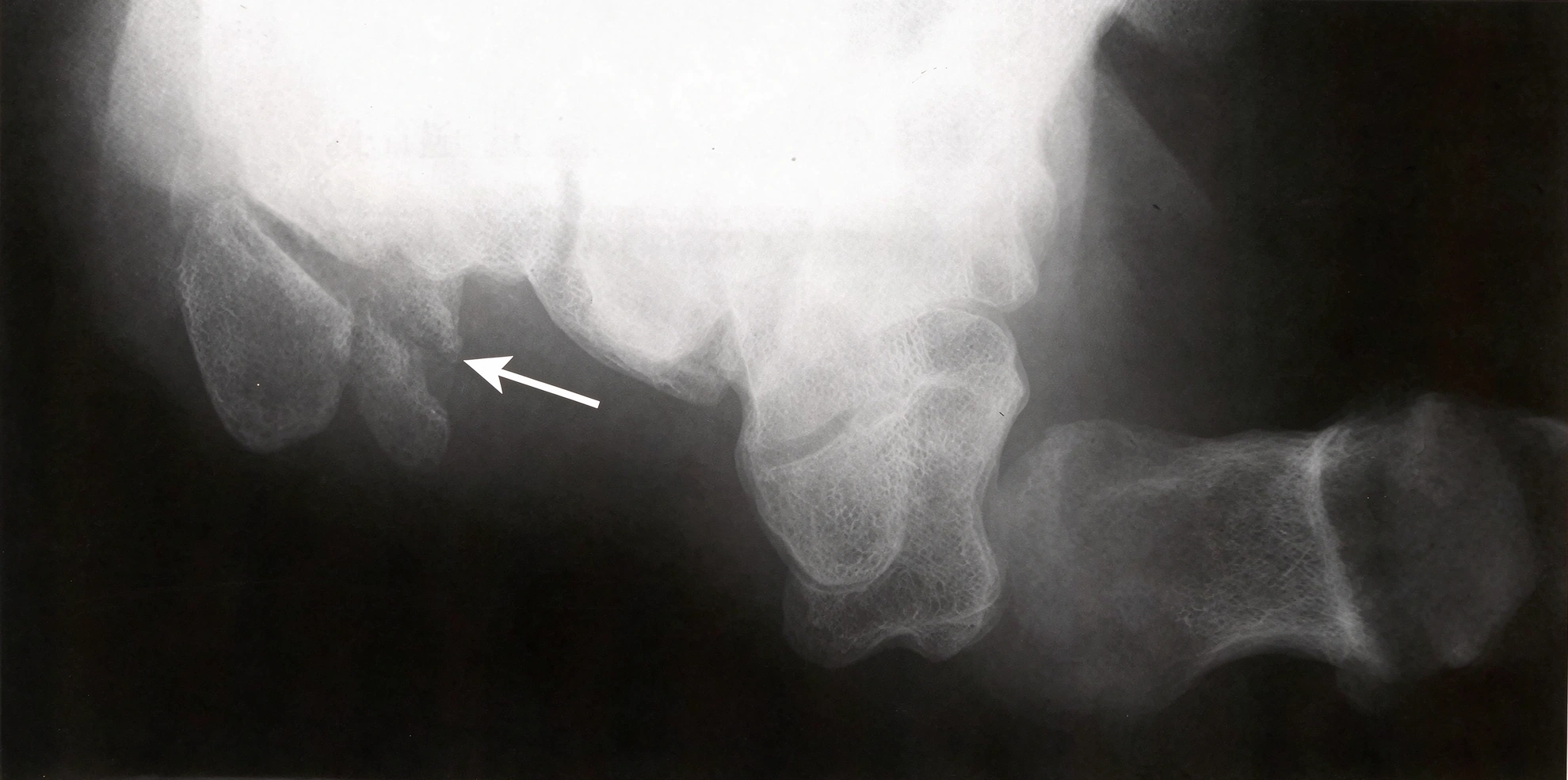
▲ The carpal tunnel view shows a hook of hamate fracture.
CT is generally the preferred imaging examination for confirming a suspected hook of hamate fracture.
Compared with conventional radiographs, CT provides more detailed visualization of the bone and can help identify:
The exact fracture location.
Fragment displacement.
Fracture configuration.
Chronic nonunion.
Associated bony abnormalities.
CT is also valuable for preoperative planning, particularly when the surgeon needs to evaluate the fracture fragment and surrounding carpal anatomy.
MRI may be useful when associated soft-tissue injuries are suspected.
It can help evaluate:
Bone marrow edema.
Flexor tendon abnormalities.
Ulnar nerve abnormalities.
Soft-tissue inflammation.
Associated ligament or cartilage injuries.
CT is generally more useful for defining the cortical fracture, while MRI provides additional information about surrounding soft tissues.
Treatment depends on fracture displacement, the time elapsed since injury, symptoms, associated injuries, and the patient's activity requirements.
The main treatment options include immobilization, fragment excision, and open reduction and internal fixation (ORIF).
Acute, nondisplaced hook of hamate fractures may be treated non-operatively in selected patients.
Treatment generally involves:
Immobilization with an appropriate cast or splint.
Restriction of gripping and strenuous wrist activities.
Follow-up clinical and imaging assessments.
Gradual rehabilitation after the initial healing period.
An ulnar gutter cast or other suitable immobilization method may be used, depending on the fracture and clinician preference.
A period of approximately six weeks of immobilization is commonly described, although the actual duration should be adjusted according to fracture healing and clinical progress.
Conservative treatment may be less predictable in fractures that are displaced, chronic, or associated with symptomatic nonunion.
Surgery may be considered in patients with:
Displaced hook fractures.
Symptomatic nonunion.
Persistent pain despite appropriate conservative treatment.
Ulnar nerve compression or irritation.
Flexor tendon irritation or rupture.
Functional limitations that prevent a return to sports or work.
Two main surgical strategies are used: fragment excision and open reduction and internal fixation.
Hook of Hamate Fragment Excision
Excision of the fractured hook is a commonly used treatment, particularly for chronic fractures, symptomatic nonunion, and selected displaced injuries.
The procedure removes the symptomatic bony fragment while preserving the remaining hamate body and surrounding soft-tissue structures.
Open Reduction and Internal Fixation
In selected acute fractures, particularly when the fragment is suitable for fixation, ORIF may be considered.
Fixation methods may include small screws or Kirschner wires, depending on fragment size, fracture configuration, and bone quality.
The choice between excision and fixation should be individualized. Fragment excision is not the only surgical option, and preserving the hook may be appropriate in selected patients.
Hook excision requires careful identification and protection of the ulnar neurovascular structures and adjacent flexor tendons.
The deep motor branch of the ulnar nerve is particularly important because it passes close to the base of the hook.
An appropriate palmar-ulnar approach is used to expose the hook of the hamate.
The soft tissues are carefully dissected to identify the relevant anatomical structures.
Important structures requiring protection include:
Ulnar artery.
Superficial sensory branch of the ulnar nerve.
Deep motor branch of the ulnar nerve.
Flexor tendons of the ring and little fingers.
The ulnar neurovascular bundle and the deep motor branch should be identified and protected throughout the procedure.
After adequate exposure, the surrounding soft tissues and periosteum are carefully elevated to expose the fractured hook.
The surgeon should pay particular attention to the base of the hook, where the deep motor branch of the ulnar nerve is especially vulnerable.
Careful subperiosteal dissection helps reduce the risk of injury to the adjacent nerve and tendons.
Once the fracture is clearly visualized, the hook fragment is removed using an appropriate surgical instrument, such as a small rongeur or other suitable instrument.
In chronic fractures, additional dissection or controlled bone removal may be required because of fibrous tissue and established nonunion.
The surgeon should avoid excessive traction on the ulnar nerve branches during fragment removal.
After removing the hook, the remaining bony surface is inspected.
Sharp or irregular bone edges are carefully smoothed to reduce the risk of continued irritation to the flexor tendons and surrounding neurovascular structures.
The surgical field is then inspected to ensure that no residual fragment or prominent bony edge remains.
The remaining periosteum and appropriate surrounding soft tissues are repaired over the base of the hamate when possible.
This helps provide soft-tissue coverage and reduce potential irritation of the flexor tendons and ulnar nerve.
The wound is then closed according to the surgeon's preferred technique.
Important Surgical Consideration: The deep motor branch of the ulnar nerve is located close to the base of the hook. It must be identified and protected during exposure, dissection, and bone removal.
Unlike hook fractures, hamate body fractures involve the main portion of the bone and are frequently associated with injuries to the fourth and fifth CMC joints.
These injuries may result from higher-energy trauma, direct impact, or axial loading transmitted through the fourth and fifth metacarpals.
Several fracture patterns have been described.
This pattern involves the proximal portion of the hamate body.
Depending on the fracture configuration, associated carpal instability or other wrist injuries may be present.
This pattern involves the medial portion of the hamate body.
The extent of displacement and involvement of adjacent articular surfaces should be assessed when determining treatment.
An oblique fracture line passes through the hamate body in a sagittal orientation.
This pattern may affect the articular surface supporting the fourth and fifth metacarpal bases and can be associated with CMC joint instability.
This pattern involves the dorsal portion of the hamate body in the coronal plane.
It may occur with dorsal fracture-dislocation of the fourth and fifth CMC joints.
CT is particularly useful for defining the fracture pattern and evaluating the degree of articular involvement.
Classification Note: Hamate body fractures have been described using several classification systems. The four patterns above provide an anatomical description of commonly discussed fracture configurations; they should not be regarded as a single universally accepted classification system.
The fourth and fifth CMC joints are important for normal hand function, particularly during gripping and adapting the hand to different object shapes.
The fourth and fifth metacarpal bases articulate with the hamate and allow a degree of movement that contributes to the flexibility of the ulnar side of the hand.
This mobility helps the hand conform to objects during power grip and improves functional grasping ability.
A hamate body fracture may disrupt the articular surfaces of these joints and compromise their stability.
Associated injuries may include:
Fourth metacarpal base fracture.
Fifth metacarpal base fracture.
Fourth CMC joint subluxation or dislocation.
Fifth CMC joint subluxation or dislocation.
Combined fourth and fifth CMC fracture-dislocation.
Failure to restore the normal alignment of these joints may lead to persistent pain, reduced grip strength, and post-traumatic arthritis.
Treatment depends on fracture displacement, stability, articular involvement, and associated CMC joint injuries.
Isolated, nondisplaced, and stable hamate body fractures may be treated with immobilization.
A cast or splint is used to protect the fracture and maintain alignment during healing.
A period of approximately six weeks of immobilization is commonly described, with follow-up imaging used to assess fracture position and healing.
Conservative treatment is generally more suitable when the fracture does not cause significant articular displacement or CMC joint instability.
Surgical treatment is generally considered when the fracture is displaced, unstable, or associated with fourth or fifth CMC joint subluxation or dislocation.
The primary objectives are to:
Restore the normal anatomy of the hamate.
Re-establish the articular alignment of the CMC joints.
Restore stability of the fourth and fifth metacarpal bases.
Maintain appropriate hand function and grip strength.
Depending on the fracture configuration, treatment may involve closed reduction and percutaneous pinning or open reduction and internal fixation.
A dorsal approach is commonly used to expose the hamate body.
The surgical interval is developed between the extensor tendons of the fourth and fifth digits, with careful protection of the surrounding soft tissues.
The fracture site and the adjacent fourth and fifth CMC joints are exposed to evaluate the extent of displacement and joint instability.
The fracture fragments are carefully reduced to restore the normal anatomical alignment of the hamate.
The fourth and fifth CMC joints should also be assessed because instability or dislocation frequently accompanies hamate body fractures.
Once the fracture and joint alignment have been restored, temporary fixation may be used to maintain the reduction.
Kirschner wires may be used to stabilize the reduced CMC joints and maintain fracture alignment.
In selected cases, wires are inserted from the affected metacarpal bases into the adjacent carpal bones to stabilize the fourth and fifth CMC joints.
The fixation configuration depends on the fracture pattern and the degree of joint instability.
Small screws, including suitable compression screws, may be used to stabilize selected hamate body fracture patterns.
Screw fixation is particularly useful when the fracture configuration allows adequate fragment purchase and compression.
The surgeon should select the screw size and trajectory according to the available bone stock and the location of adjacent anatomical structures.
A small, low-profile plate, including an appropriately selected mini-plate, may be used when additional fixation stability is required.
Plate fixation may be helpful in more complex or comminuted fracture patterns where screw fixation alone is insufficient.
The choice of fixation method should be based on fracture morphology, articular involvement, bone quality, and the need to restore CMC joint stability.
Important Surgical Consideration: The deep motor branch of the ulnar nerve lies close to the palmar aspect of the hamate. Particular care is required when drilling from dorsal to palmar, selecting screw length, or placing fixation devices to avoid nerve injury.
Delayed diagnosis or inadequate treatment may result in several complications.
Nonunion
The fracture may fail to achieve bony union, particularly in chronic hook fractures.
Ulnar Nerve Injury
The fracture or surgical procedure may irritate or injure the ulnar nerve or its branches, resulting in sensory changes or intrinsic muscle weakness.
Flexor Tendon Rupture
Chronic irritation from a hook fracture fragment may cause partial or complete rupture of the flexor tendons of the ring and little fingers.
Reduced Grip Strength
Persistent pain, nerve dysfunction, tendon injury, or CMC joint incongruity may reduce grip strength.
Post-Traumatic Arthritis
Inadequately reduced hamate body fractures involving the CMC articular surfaces may lead to post-traumatic joint degeneration.
Persistent CMC Joint Instability
Failure to restore stable alignment of the fourth and fifth CMC joints may result in chronic instability and functional impairment.
Q1: What is the difference between a hook of hamate fracture and a hamate body fracture?
A hook of hamate fracture involves the palmar hook-shaped projection of the bone and is commonly associated with gripping injuries. A hamate body fracture involves the main portion of the bone and may be associated with fourth and fifth CMC joint fracture-dislocations.
Q2: Why are hook of hamate fractures difficult to diagnose?
The hook is often obscured by overlapping carpal bones on standard wrist X-rays. Special radiographic views may help, but CT is generally preferred when a fracture is suspected.
Q3: Can a hook of hamate fracture heal without surgery?
Yes. Selected acute, nondisplaced fractures may heal with appropriate immobilization. However, displaced fractures, symptomatic nonunion, persistent pain, and associated tendon or nerve injuries may require surgery.
Q4: Why can a hook of hamate fracture cause numbness in the ring and little fingers?
The ulnar nerve passes close to the hook of the hamate through Guyon's canal. A fracture fragment or surrounding inflammation may irritate or compress the nerve, causing sensory symptoms or muscle weakness.
Q5: What is the Milch classification of hook of hamate fractures?
The Milch classification describes three fracture locations: the tip, waist, and base of the hook of the hamate.
Q6: When does a hamate body fracture require surgery?
Surgery may be necessary when the fracture is displaced or unstable, involves important articular surfaces, or is associated with fourth or fifth CMC joint subluxation or dislocation.
Q7: What fixation methods are used for hamate body fractures?
Depending on fracture morphology, fixation may involve Kirschner wires, small compression screws, or low-profile plates. The goal is to restore anatomical alignment and maintain stable fourth and fifth CMC joints.
Hamate fractures are uncommon carpal injuries that require careful clinical and radiographic evaluation. They are broadly divided into hook fractures and body fractures, each with distinct anatomical characteristics and treatment considerations.
Hook of hamate fractures may cause hypothenar pain, reduced grip strength, ulnar nerve symptoms, and flexor tendon irritation. Because standard radiographs may fail to identify these injuries, CT is particularly valuable for confirming the diagnosis and defining the fracture pattern.
Hamate body fractures frequently involve the fourth and fifth CMC joints. Stable, nondisplaced injuries may be managed with immobilization, while displaced or unstable fractures may require reduction and internal fixation using Kirschner wires, screws, or plates.
Early diagnosis, appropriate treatment selection, accurate fracture reduction, and careful protection of the ulnar nerve and flexor tendons are essential for restoring hand function and minimizing complications.


