
Views: 0 Author: Site Editor Publish Time: 2026-11-16 Origin: Site
Chronic ankle instability (CAI) is a common condition that develops when the ankle repeatedly gives way, feels weak, or lacks stability during walking, exercise, or sports activities. It often occurs after one or more ankle sprains, particularly when the injured ligaments do not recover adequately or when rehabilitation is incomplete.
Ankle sprains are among the most frequent musculoskeletal injuries. Some patients continue to experience recurrent sprains, persistent swelling, pain, and a feeling of instability after the initial injury. Without appropriate treatment, these symptoms may interfere with daily activities and athletic performance.
Understanding the anatomy of the ankle, the mechanisms of ligament injury, and the available treatment options is essential for preventing recurrent injuries and maintaining long-term joint function.
Chronic ankle instability refers to a condition in which the ankle repeatedly gives way or feels unstable following an initial ankle sprain or other ligament injury.
Patients may describe their ankle as:
Weak or lacking strength
Loose or unstable
Likely to roll or give way when walking
Unreliable when running, jumping, or changing direction
Chronic ankle instability commonly develops after an injury to the lateral ankle ligaments. These ligaments help maintain the normal position of the ankle joint and prevent excessive movement.
Following a severe ankle sprain, some patients experience persistent ligament laxity, impaired balance, reduced muscle coordination, or altered joint position awareness. These problems can contribute to repeated ankle sprains.
Research has reported that a substantial proportion of patients with significant ankle sprains develop recurrent symptoms or chronic instability. However, the likelihood varies depending on injury severity, rehabilitation, activity level, and individual risk factors.
The ankle is a complex joint connecting the lower leg to the foot. It plays an essential role in supporting body weight, maintaining balance, and allowing movements such as walking, running, jumping, and turning.
The ankle joint is primarily formed by three bones:
Tibia: The larger bone of the lower leg, forming the medial portion of the ankle joint.
Fibula: The smaller bone on the outer side of the lower leg, forming the lateral ankle.
Talus: The bone positioned between the tibia, fibula, and heel, allowing ankle movement.
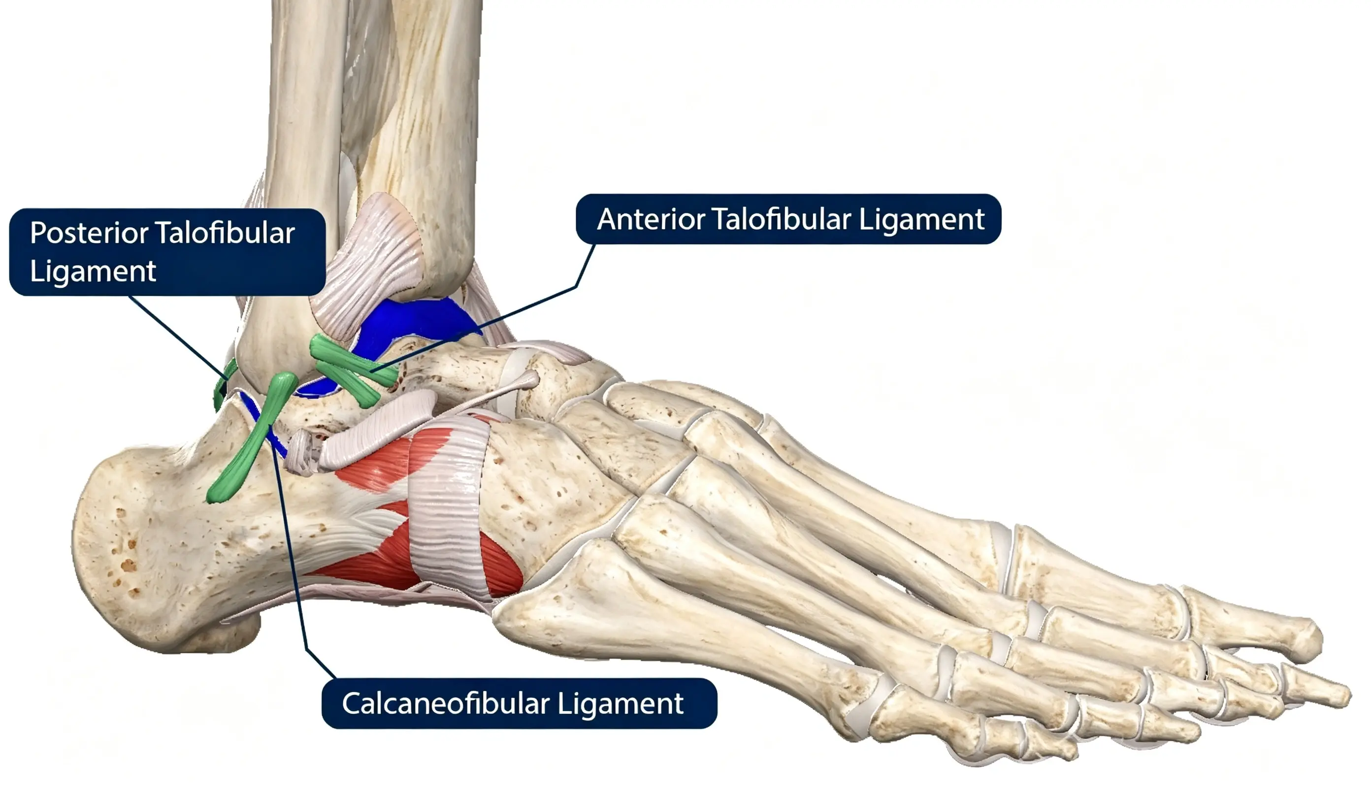
Ligaments are strong bands of fibrous connective tissue that connect bones and help stabilize joints.
The lateral side of the ankle contains three major ligaments:
1. Anterior Talofibular Ligament (ATFL)
The ATFL connects the fibula to the talus at the front of the lateral ankle. It is the most frequently injured ligament during an inversion ankle sprain, particularly when the foot rolls inward.
2. Calcaneofibular Ligament (CFL)
The CFL connects the fibula to the calcaneus, or heel bone. It contributes to lateral ankle stability and may be injured in moderate or severe ankle sprains.
3. Posterior Talofibular Ligament (PTFL)
The PTFL connects the fibula to the posterior part of the talus. It is relatively strong and is less commonly injured than the ATFL and CFL. Injury to this ligament is more often associated with severe ankle trauma.
Among these structures, the ATFL is the most commonly affected ligament in lateral ankle sprains. Combined ATFL and CFL injuries may occur in more severe cases.
An ankle sprain occurs when one or more ligaments are stretched beyond their normal limits or partially or completely torn.
Most lateral ankle sprains happen when the foot suddenly rolls inward, a movement known as inversion. This places excessive stress on the lateral ankle ligaments.
Ankle sprains are commonly classified into three grades according to the severity of ligament damage.
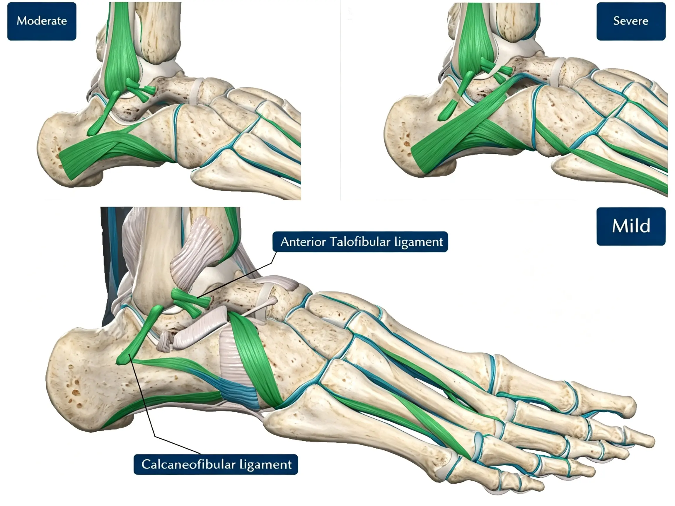
The ligament is mildly stretched.
There may be minor microscopic fiber damage.
Swelling and tenderness are usually limited.
The ankle generally remains stable.
Recovery is often relatively quick with appropriate treatment.
The ligament is partially torn.
Pain, swelling, and bruising are more noticeable.
Some joint instability may be present.
Walking and physical activities may be difficult.
The ligament is completely torn.
Significant swelling and bruising may occur.
The ankle may demonstrate substantial instability.
Weight-bearing and sports activities may be severely restricted.
In severe injuries, damage may involve more than one ligament. Cartilage injuries, bone bruising, or associated fractures may also occur.
Clinical Note: The severity of an ankle sprain cannot always be determined by pain or swelling alone. A proper clinical examination and, when indicated, imaging studies are important for evaluating the extent of the injury.
Chronic ankle instability most commonly develops following an ankle sprain that does not recover adequately.
Several factors may contribute to persistent instability.
After an ankle sprain, damaged ligaments undergo a healing process. In some patients, the ligaments may heal in an elongated or mechanically weakened position.
This can reduce the ability of the lateral ankle ligaments to control excessive joint movement.
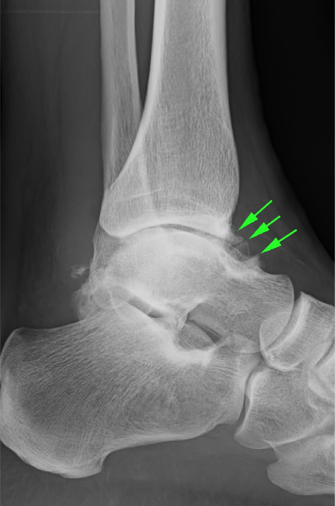
▲ Chronic ankle instability with X-ray showing osteophytes at the anterior joint margin (green arrow).
Rest alone may not restore normal ankle function. Even after pain and swelling improve, patients may continue to experience:
Reduced ankle muscle strength
Poor balance and coordination
Impaired proprioception
Delayed muscular responses
Reduced functional stability
Without appropriate rehabilitation, these problems may increase the risk of recurrent ankle sprains.
Patients who experience repeated inversion injuries may develop progressive ligament damage and persistent joint instability.
Each new sprain can further affect ligament function, balance, and confidence during physical activity.
Proprioception refers to the body's ability to recognize joint position and movement without relying entirely on vision.
After an ankle injury, proprioceptive function and the coordination of surrounding muscles may become impaired. This can make it harder for patients to react quickly when the ankle begins to roll.
Ankle sprains are among the most common injuries in sports and everyday activities.
They frequently occur in sports involving jumping, running, sudden acceleration, and rapid changes in direction, including:
Soccer
Basketball
Tennis
Volleyball
Rugby
Trail running
Ankle sprains can also occur during ordinary activities, such as walking on uneven surfaces, exercising, running outdoors, or missing a step on a staircase.
Athletes with a history of ankle sprains may have a higher risk of recurrent injuries, particularly when they return to sports before restoring adequate strength, balance, and functional stability.
The symptoms of chronic ankle instability may develop gradually after an initial ankle sprain.
Common symptoms include:
Repeated giving-way episodes: The ankle suddenly feels as though it is rolling or collapsing during movement.
Recurrent ankle sprains: Patients experience repeated inversion injuries, sometimes during relatively minor activities.
Pain: Discomfort may occur around the outer ankle, especially after exercise or prolonged walking.
Swelling: Persistent or intermittent swelling may develop around the ankle joint.
Stiffness: Reduced ankle mobility may make walking, squatting, or running uncomfortable.
Weakness: The ankle may feel weak or unreliable during physical activities.
Reduced balance: Patients may have difficulty standing on one leg or moving confidently on uneven surfaces.
Not every patient with chronic ankle instability experiences severe pain.
Some patients mainly report repeated giving-way episodes, while others experience persistent discomfort and swelling.
Pain may be associated with several conditions, including:
Recurrent ligament injuries
Synovitis or inflammation of the joint lining
Anterolateral or anteromedial ankle impingement
Osteochondral lesions of the talus
Peroneal tendon injuries
Post-traumatic cartilage and joint damage
In particular, an osteochondral lesion may develop following an ankle injury. Because articular cartilage has limited direct nerve supply, cartilage damage may not always produce obvious symptoms initially. However, associated damage to the underlying subchondral bone and surrounding tissues can cause pain.
Repeated joint trauma may also contribute to the development of osteophytes, commonly called bone spurs. These bony changes can restrict ankle movement and contribute to joint impingement.
Yes. Chronic ankle instability should not be ignored, especially when patients experience repeated sprains, persistent pain, or difficulty with daily activities.
Potential complications include:
Repeated ankle sprains can cause further damage to the lateral ligaments and surrounding soft tissues.
Ankle trauma may damage the cartilage and underlying bone of the talus. These lesions can cause persistent pain, swelling, and mechanical symptoms.
Repeated inflammation and joint trauma may contribute to soft-tissue thickening or osteophyte formation. These changes can cause pain and restricted movement when the ankle is brought into certain positions.
Chronic instability and repeated joint injuries may increase the risk of post-traumatic ankle osteoarthritis.
However, ankle instability does not inevitably lead to arthritis. The risk depends on factors such as the severity of the original injury, associated cartilage damage, joint alignment, and the frequency of recurrent trauma.
In advanced cases of ankle arthritis, patients may require surgical treatment such as ankle arthrodesis or total ankle replacement.
Ankle Arthrodesis: This procedure permanently fuses the ankle joint to reduce pain caused by joint movement. It can provide reliable pain relief in selected patients, but ankle motion is sacrificed.
Total Ankle Replacement: This procedure replaces the damaged joint surfaces with artificial components to relieve pain while preserving some ankle movement. Patient selection and implant-related considerations are important.
These procedures are treatments for selected cases of advanced ankle arthritis, rather than routine treatments for chronic ankle instability alone.
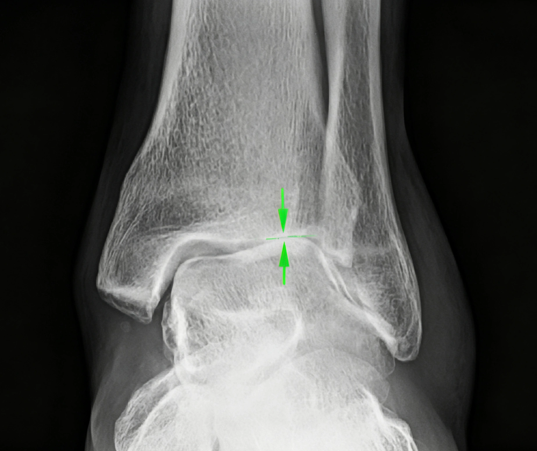
▲ X-ray of the foot and ankle joint in a patient with arthritis. (Green arrows indicate areas of cartilage loss and osteophyte formation.)
Chronic ankle instability may be overlooked because symptoms can develop gradually. Some patients assume that repeated ankle weakness or occasional giving-way episodes are simply the result of a previous sprain.
An accurate diagnosis requires a combination of medical history, physical examination, and appropriate imaging.
The doctor may ask about:
Previous ankle sprains and their severity
The number of recurrent ankle injuries
Episodes of the ankle giving way
Pain and swelling patterns
Sports and daily activities that trigger symptoms
Previous rehabilitation and treatment
Any history of fractures or other ankle injuries
The doctor will examine the ankle to identify possible structural and functional abnormalities.
Important assessment points include:
Tenderness: Identifying painful areas around the lateral ankle ligaments and joint.
Swelling: Checking for persistent or recurrent swelling.
Ligament laxity: Evaluating whether the ankle ligaments are excessively loose.
Anterior drawer test: Assessing the integrity of the ATFL.
Talar tilt test: Evaluating lateral ligament stability, particularly the CFL.
Range of motion: Checking for restricted movement or joint stiffness.
Impingement: Looking for signs of soft-tissue or bony impingement.
Balance and functional stability: Evaluating single-leg balance and movement control.
Imaging studies may be recommended depending on the patient's symptoms and examination findings.
X-rays
X-rays help identify fractures, joint alignment abnormalities, osteophytes, and signs of ankle osteoarthritis.
Magnetic Resonance Imaging (MRI)
MRI can help evaluate the ATFL, CFL, joint cartilage, osteochondral lesions, tendons, and other soft-tissue structures.
Ultrasound
Musculoskeletal ultrasound can be used to assess superficial ankle ligaments and may help identify ligament abnormalities during dynamic examination.
Stress Radiographs
In selected patients, stress radiographs can help evaluate mechanical ankle instability by demonstrating abnormal joint movement under applied stress.
Not every patient requires all these examinations. The choice of imaging depends on the suspected injury and whether the results will affect treatment decisions.
Treatment depends on the severity of instability, the patient's activity level, associated injuries, and response to previous treatment.
In most cases, non-surgical treatment is the first-line approach.
The main objectives are to:
Reduce pain and swelling
Restore ankle range of motion
Improve muscle strength
Restore balance and proprioception
Improve neuromuscular control
Prevent recurrent ankle sprains
For an acute ankle sprain, treatment may include activity modification, protection of the injured ankle, compression, elevation, and appropriate pain management.
The traditional RICE approach includes:
Rest: Temporarily reduce activities that aggravate the injury.
Ice: Apply cold therapy for short periods to help manage pain.
Compression: Use an appropriate compression bandage to help control swelling.
Elevation: Raise the injured ankle to help reduce swelling.
Depending on injury severity, a brace or short-term immobilization may also be recommended.
Patients should avoid prolonged unnecessary immobilization when it is not clinically indicated. Early protected movement and progressive weight-bearing are often incorporated into rehabilitation as tolerated.
Rehabilitation is one of the most important components of treating chronic ankle instability.
A personalized rehabilitation program may include:
1. Range-of-Motion Exercises
Exercises help restore ankle flexibility and reduce stiffness following injury.
2. Muscle Strengthening
Strengthening the peroneal muscles, calf muscles, and other supporting muscle groups can improve dynamic ankle stability.
3. Balance Training
Single-leg standing, balance-board exercises, and controlled weight-shifting activities help improve postural control.
4. Proprioceptive Training
These exercises improve the body's ability to recognize ankle position and respond to unexpected joint movement.
5. Neuromuscular Training
Progressive movement exercises help improve coordination and the speed of muscular responses during walking, running, jumping, and changes of direction.
6. Functional and Sports-Specific Training
Patients returning to sports may need progressive exercises involving jumping, landing, acceleration, deceleration, and directional changes.
An ankle brace or taping may also be considered during higher-risk activities, particularly for patients with a history of recurrent sprains.
There is no single rehabilitation duration that is appropriate for every patient.
A structured physical therapy program commonly lasts at least 6 weeks to 3 months. Some patients, particularly those with persistent weakness, poor balance, or demanding sports requirements, may need rehabilitation for up to 6 months.
Treatment duration should depend on functional improvement rather than time alone.
Before returning to high-demand sports, patients should demonstrate adequate:
Ankle strength
Range of motion
Balance and proprioception
Single-leg control
Jumping and landing ability
Confidence during sports-specific movements
If instability persists despite an appropriate rehabilitation program, further clinical assessment may be necessary.
Most patients initially receive non-surgical treatment. However, some continue to experience recurrent ankle sprains, mechanical instability, or functional limitations despite adequate rehabilitation.
Surgery may be considered when patients have:
Repeated ankle sprains or giving-way episodes
Persistent mechanical instability confirmed by physical examination
Symptoms that interfere with daily activities or sports
Inadequate improvement after structured rehabilitation
Associated injuries requiring surgical treatment
The decision to operate should be based on the patient's symptoms, physical examination, imaging findings, activity demands, and response to conservative treatment.
For patients with persistent lateral ankle instability, surgical treatment commonly focuses on restoring the function of the damaged lateral ligaments.
Anatomic Ligament Repair
The modified Broström procedure is a commonly used surgical technique for chronic lateral ankle instability.
It involves repairing and tightening the damaged lateral ankle ligaments, particularly the ATFL, and may include the CFL depending on the injury pattern.
The goal is to restore the normal anatomy and mechanical stability of the ankle while preserving joint movement.
Ligament Reconstruction
In patients with poor ligament tissue quality, significant chronic laxity, failed previous repair, or selected high-demand cases, ligament reconstruction may be considered.
This procedure uses a tendon graft to reconstruct the damaged ligament structures and restore ankle stability.
The choice between ligament repair and reconstruction depends on ligament quality, injury characteristics, patient factors, and the surgeon's assessment.
Treatment of Associated Pathology
If chronic ankle instability is accompanied by osteochondral lesions, synovitis, or bony impingement, these conditions may also need to be addressed during treatment.
Q1: Can chronic ankle instability heal without surgery?
Yes. Many patients improve with structured physical therapy, muscle strengthening, balance training, proprioceptive exercises, and appropriate activity modification. Surgery is generally considered when symptoms persist despite adequate conservative treatment.
Q2: Why does my ankle keep rolling after a sprain?
Repeated ankle rolling may result from ligament laxity, incomplete ligament healing, reduced muscle strength, impaired proprioception, or poor neuromuscular control.
Q3: How long does it take to recover from chronic ankle instability?
Recovery varies depending on injury severity, rehabilitation progress, and activity requirements. Some patients improve within several weeks to a few months, while others require longer rehabilitation or surgical treatment.
Q4: Can I continue playing sports with chronic ankle instability?
Some patients can continue sports with appropriate rehabilitation, strengthening, and ankle support. However, repeated giving-way episodes increase the risk of further injury. Returning to sports should be based on functional recovery rather than pain reduction alone.
Q5: Does chronic ankle instability always cause ankle arthritis?
No. Although repeated ankle injuries and persistent instability may increase the risk of post-traumatic osteoarthritis, not every patient develops arthritis. Early diagnosis and appropriate management may help reduce further joint damage.
Q6: When should I consider ankle instability surgery?
Surgery may be considered when recurrent sprains, mechanical instability, or functional limitations continue despite an adequate course of non-surgical treatment. A foot and ankle specialist can evaluate whether ligament repair or reconstruction is appropriate.
Chronic ankle instability is a common long-term complication of ankle sprains, particularly when ligament healing and rehabilitation are inadequate. Patients may experience recurrent ankle sprains, weakness, swelling, pain, and repeated episodes of the ankle giving way.
Early diagnosis and structured rehabilitation are essential for restoring ankle strength, balance, proprioception, and functional stability. For patients who continue to experience significant instability despite appropriate non-surgical treatment, surgical ligament repair or reconstruction may provide another treatment option.
Recognizing the symptoms and addressing the underlying causes can help reduce recurrent injuries, improve ankle function, and support a safer return to daily activities and sports.


