
Views: 0 Author: Site Editor Publish Time: 2026-11-18 Origin: Site
Not every visible deviation of the spine indicates scoliosis. Many people have some degree of postural asymmetry or spinal deviation from the body's midline. However, a clinical diagnosis of scoliosis requires more than simply observing that the spine is not perfectly straight.
Scoliosis is a three-dimensional spinal deformity involving a lateral curvature, often accompanied by vertebral rotation. It is generally defined radiographically by a Cobb angle of at least 10 degrees.
One of the most widely used clinical screening methods is the Adams Forward Bend Test, which helps identify trunk asymmetry and rotational prominence. By observing the patient's back in different positions, clinicians can obtain useful information about the possible presence and characteristics of scoliosis.
This article explains the differences between functional and structural scoliosis, how to perform the Adams test, why rib prominence occurs, and how to document postural changes during treatment.
Scoliosis can be classified in several ways, including by cause, age of onset, curve location, and structural characteristics.
From a clinical assessment perspective, an important distinction is between functional (nonstructural) scoliosis and structural scoliosis.
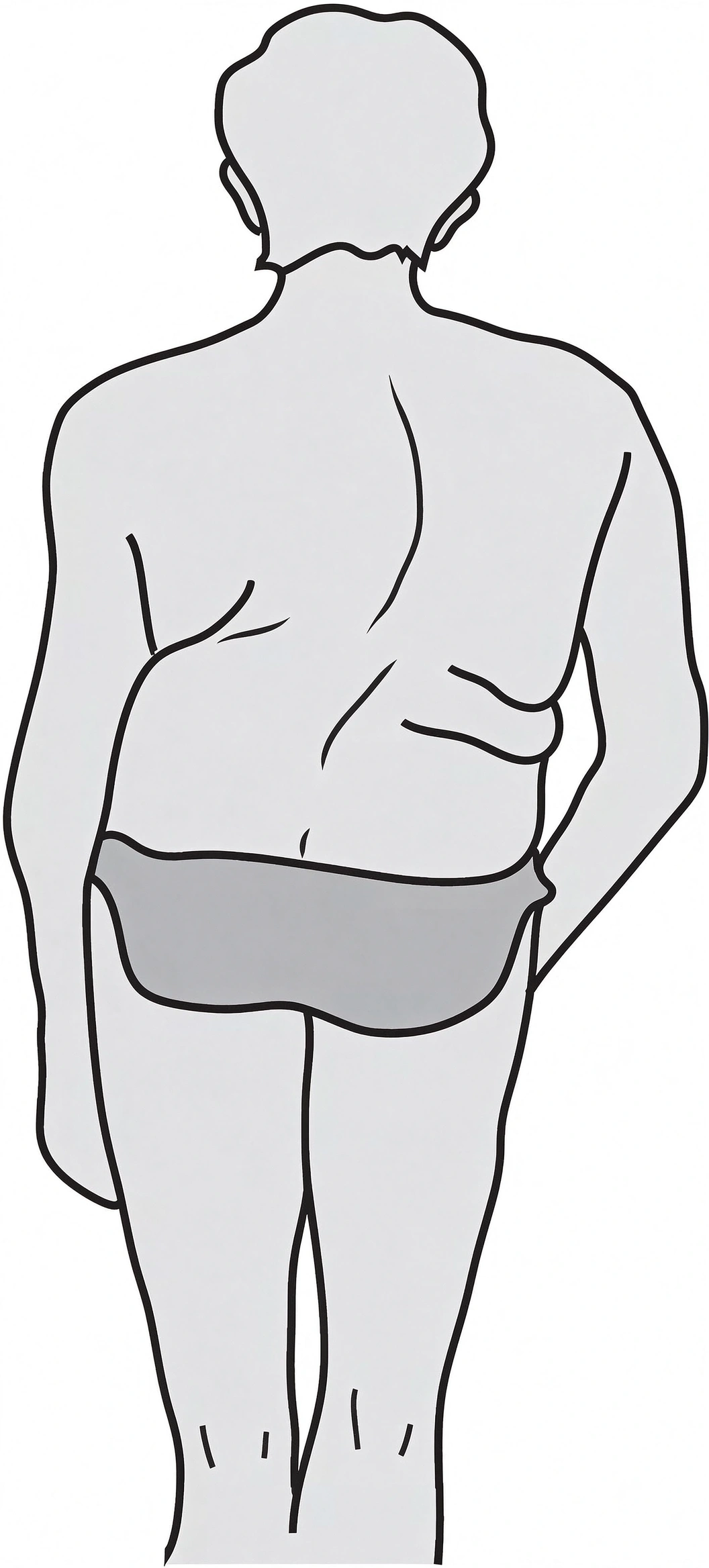
Functional scoliosis, sometimes called flexible or nonstructural scoliosis, refers to a lateral spinal curve that develops in response to an underlying factor rather than a fixed structural deformity of the vertebrae.
Possible contributing factors include:
Leg-length discrepancy
Pelvic obliquity
Muscle spasm or pain-related postural compensation
Habitual asymmetric posture
Other conditions that cause the patient to adopt an uneven standing position
The curve may become smaller or disappear when the underlying cause is corrected or when the patient changes position.
For example, a patient with a functional curve related to pelvic obliquity may demonstrate improved spinal alignment when the pelvis is leveled.
However, the degree of correction varies depending on the underlying cause. Functional scoliosis should not automatically be attributed to muscle or ligament abnormalities alone.
Structural scoliosis involves a more fixed spinal deformity, typically with vertebral rotation and changes in the normal alignment of the spine.
Unlike a flexible postural curve, a structural curve generally does not disappear simply because the patient lies down or changes position.
Common categories include:
Idiopathic scoliosis
Congenital scoliosis
Neuromuscular scoliosis
Degenerative scoliosis
Structural scoliosis may produce visible asymmetry of the shoulders, shoulder blades, waist, pelvis, and rib cage.
Vertebral rotation is particularly important because it can create a rib prominence on one side of the back, which may be detected during the Adams Forward Bend Test.
| Assessment Feature | Functional Scoliosis | Structural Scoliosis |
| Underlying characteristics | Usually related to a reversible postural or mechanical factor | Involves a more fixed spinal deformity |
| Spinal flexibility | Generally more flexible | Usually less flexible |
| Adams Forward Bend Test | Trunk asymmetry may decrease or be less prominent | Rotational prominence often remains visible |
| Supine positioning | The curve may decrease or correct | The structural curve generally persists |
| Voluntary correction | May improve with posture or correction of the underlying cause | Usually cannot be completely corrected voluntarily |
| Vertebral rotation | Usually absent or limited | Commonly present |
Important: These findings are clinical clues rather than definitive diagnostic criteria. A structural curve may show some flexibility, while a functional curve may not completely disappear in every position. Clinicians may use side-bending radiographs and other assessments to evaluate curve flexibility.
Adams (1820–1900) is associated with the clinical examination commonly known as the Adams Forward Bend Test.
The test is widely used in scoliosis screening because forward bending makes certain forms of trunk asymmetry and vertebral rotation easier to observe.
During the examination, the clinician looks for:
Rib prominence on one side of the back
Unevenness of the thoracic region
Asymmetry of the lumbar or flank region
Differences in the contour of the back
A raised area on one side of the back may indicate rotational deformity associated with scoliosis.
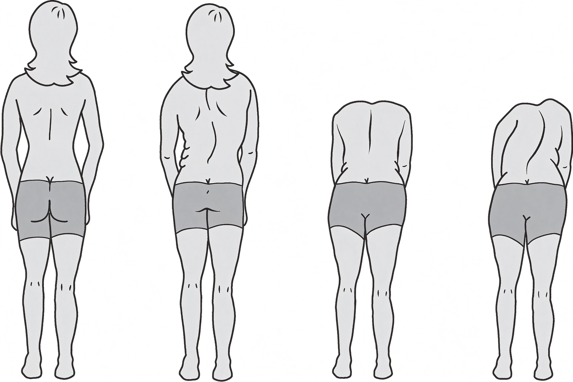
Patient position:
Ask the patient to stand upright with the feet together.
Keep the knees straight and allow the arms to hang naturally.
Ask the patient to bend forward slowly from the waist.
The patient should allow the head and arms to relax toward the floor.
Continue bending until the trunk is approximately horizontal, as tolerated.
Examiner position and observation:
Stand behind the patient and observe the back.
Compare the height and contour of both sides of the rib cage.
Look for a raised area on one side of the thoracic spine.
Observe the lumbar region for asymmetric paraspinal or flank prominence.
If available, use a scoliometer to measure the angle of trunk rotation.
Interpretation:
A visible rib hump or asymmetric trunk prominence is considered a positive screening finding that warrants further assessment.
The raised side often corresponds to the convex side of the thoracic curve, but the test does not independently establish the exact curve pattern or Cobb angle.
In a flexible, nonstructural curve, the visible lateral deviation may decrease when the patient bends forward or changes position.
In structural scoliosis, the rotational component often remains visible during forward bending. A rib hump or lumbar prominence may therefore become easier to identify.
The patient's position can also provide useful information:
During forward bending: A structural rotational prominence may remain visible or become more apparent.
In the supine position: A functional curve may decrease, while a structural curve generally remains.
During voluntary correction: A flexible curve may improve, whereas a structural curve is usually more difficult to correct completely.
These observations help guide further examination, but they should not be used alone to classify or diagnose scoliosis.
To understand the Adams test, it is helpful to understand the relationship between vertebral rotation and the rib cage.
Scoliosis is not simply a sideways curve. In many structural curves, the vertebrae also rotate around their vertical axes.
Because the ribs attach to the thoracic vertebrae, vertebral rotation changes the position and orientation of the ribs. This produces an asymmetric appearance of the chest wall.
For example, in a typical right-convex thoracic curve:
The thoracic vertebral bodies rotate toward the convex side.
The spinous processes rotate in the opposite direction.
The ribs on the convex side may become more prominent posteriorly.
The ribs on the opposite side may appear more prominent anteriorly.
This three-dimensional change in the rib cage is responsible for the characteristic rib hump that can be observed during forward bending.
The following table illustrates the typical rotational pattern described in a simplified thoracic scoliosis model.
| Feature | Left-Convex Thoracic Curve | Right-Convex Thoracic Curve |
| Curve convexity | Left side | Right side |
| Vertebral body rotation | Commonly toward the left | Commonly toward the right |
| Spinous process direction | Generally toward the right | Generally toward the left |
| Posterior rib prominence | Left side | Right side |
| Anterior rib prominence | Right side | Left side |
| Rib cage appearance | Asymmetric chest contour | Asymmetric chest contour |
The exact rotational pattern depends on the curve location and configuration. Double curves, thoracolumbar curves, and lumbar curves may produce different patterns of trunk asymmetry.
Clinical tip: When performing the Adams test, examine both the thoracic and lumbar regions. A patient may have a thoracic rib prominence, a lumbar prominence, or more than one area of rotational asymmetry.
Question: How can you assess whether a treatment intervention has improved spinal alignment?
There are several ways to monitor changes in scoliosis and posture. One simple clinical approach is to use standardized photographs and consistent anatomical landmarks.
This method can help document changes in visible posture before and after an intervention.
The following landmarks can be identified and marked before taking photographs:
C7 spinous process: The prominent spinous process at the base of the neck.
L4 spinous process: A lumbar spinal landmark that can help document lower trunk alignment when identified accurately.
Superior angle of the scapula: The upper medial region of the shoulder blade.
Inferior angle of the scapula: The lower tip of the shoulder blade.
Acromion: The lateral bony prominence of the shoulder.
Posterior superior iliac spine (PSIS): A palpable landmark on the posterior pelvis.
These landmarks can help compare shoulder height, scapular position, pelvic alignment, and overall trunk posture.
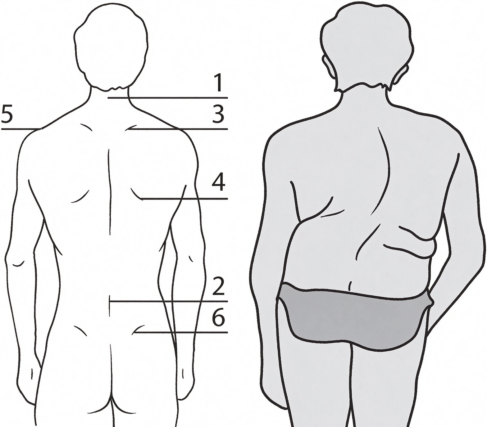
For meaningful comparison, photographs should be taken under consistent conditions.
A practical approach includes:
Use the same camera position and distance.
Maintain the same patient standing position.
Use consistent lighting and background conditions.
Photograph the patient from the posterior view.
Keep the arms and feet in the same position for each assessment.
Mark the same anatomical landmarks before each photograph.
Compare the photographs taken before and after the intervention.
This approach can help identify changes in visible body alignment, including shoulder level, scapular symmetry, trunk shift, and pelvic position.
Standardized clinical photographs provide information about changes in the relative positions of external anatomical landmarks.
For example, they may help clinicians document:
Improved shoulder symmetry
Changes in scapular position
Reduced visible trunk deviation
Changes in pelvic alignment
Improved overall postural balance
However, photographs do not directly measure the spinal Cobb angle.
If the objective is to determine whether the actual spinal curve has changed, radiographic assessment is required. The Cobb method, measured on appropriate standing spinal radiographs, remains the standard method for quantifying the magnitude of scoliosis.
Surface topography and other non-radiographic assessment methods may provide additional information for monitoring, but they should be interpreted as complementary tools rather than direct replacements for radiographic measurement.
An improvement in external posture does not necessarily mean that the structural spinal curve has been corrected.
A comprehensive evaluation should also consider the patient's functional status, symptoms, and activity level.
Depending on the clinical situation, relevant outcomes may include:
Changes in pain or discomfort
Improvement in balance and movement
Changes in daily activity performance
Respiratory function, when thoracic deformity or pulmonary impairment is a concern
Changes in spinal alignment on appropriate imaging
The choice of outcome measures should depend on the patient's age, curve type, symptoms, and treatment goals.
Question: Is the examiner's position important when performing the Adams Forward Bend Test?
The Adams test is commonly performed with the examiner standing behind the patient. This position allows the clinician to compare the two sides of the back and observe posterior rib or lumbar prominence.
However, some patients may feel uncomfortable when being examined from behind.
The examiner may also observe relevant trunk asymmetry from the front, particularly when assessing the anterior chest wall and overall body symmetry.
For a complete assessment, it is useful to observe the patient from more than one direction:
Posterior view: Assess posterior rib prominence, scapular asymmetry, and lumbar contour.
Anterior view: Observe chest wall symmetry, shoulder position, and other visible differences.
Lateral view: Assess sagittal alignment and overall posture.
The posterior view remains particularly useful for identifying the rib hump associated with vertebral rotation.
The examiner should explain the procedure, maintain appropriate patient privacy, and choose a position that allows a clear and comfortable examination.
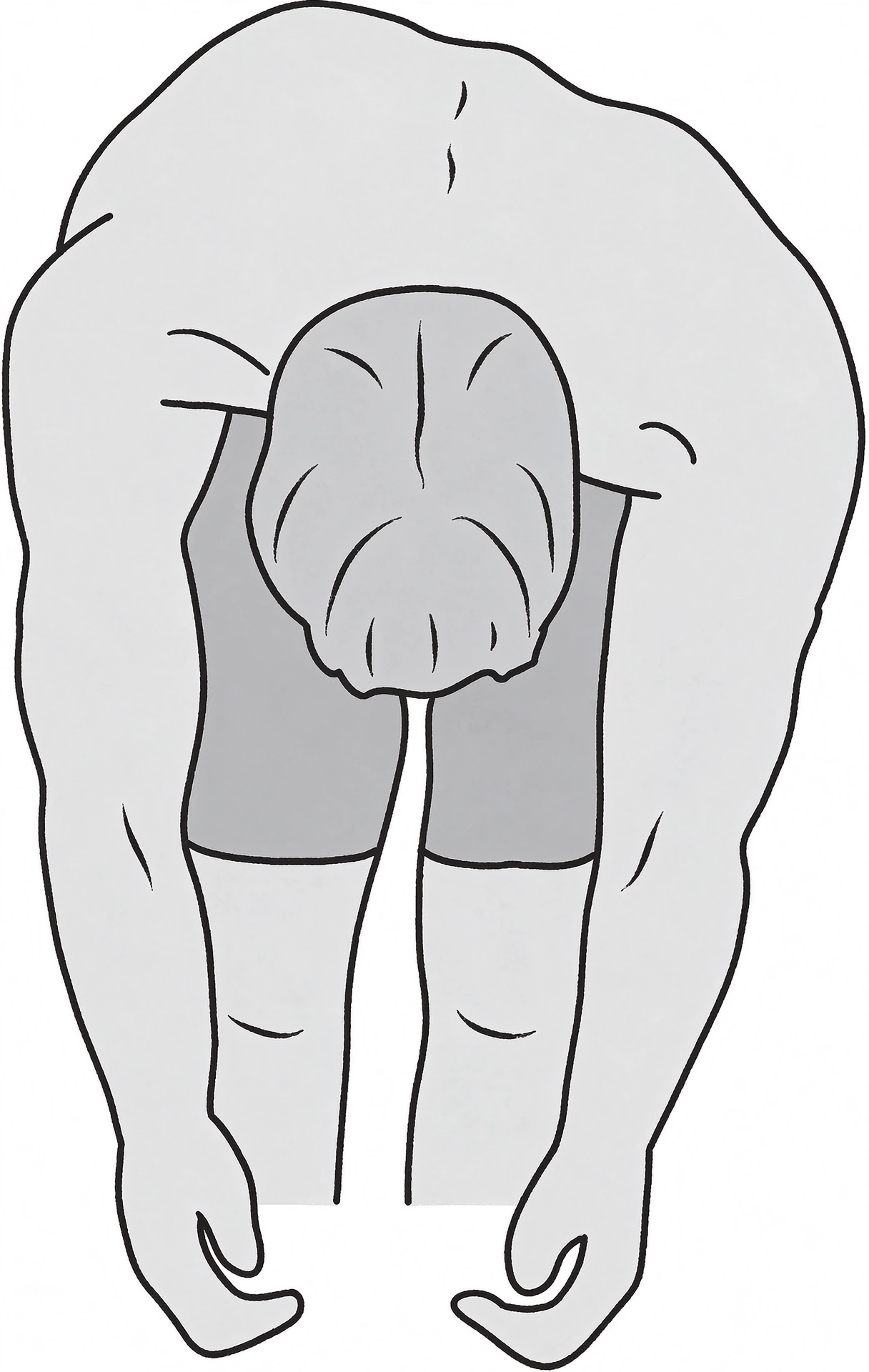
In addition to the standard scoliosis assessment methods described above, a simple visual technique has sometimes been used to obtain a quick impression of spinal alignment from the posterior side.
This technique involves using the fingernail edge as a temporary skin marker.
Ask the patient to stand upright with the back exposed.
Stand directly behind the patient.
Identify the approximate line of the spinous processes.
Gently press the side of a fingernail against the skin beside successive spinal levels.
Leave a series of small, temporary impressions without scratching or breaking the skin.
Observe the alignment of the marks from top to bottom.
Compare the resulting line with the body's approximate vertical midline.
If the marks appear to deviate noticeably from a vertical line, this may suggest visible spinal or trunk asymmetry.
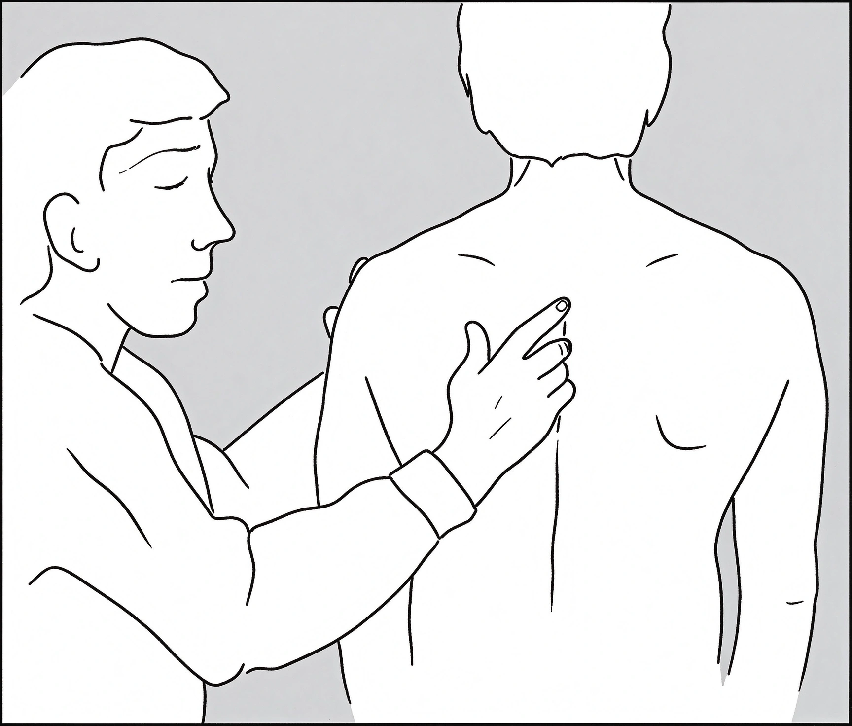
This is an informal observation technique, not a validated scoliosis screening or diagnostic test.
The skin impressions may be affected by:
Soft-tissue thickness
Difficulty identifying individual spinous processes
Patient posture and weight distribution
Examiner technique
Natural anatomical asymmetry
A line of skin marks cannot reliably establish the presence, location, or severity of scoliosis.
For this reason, the technique should never replace the Adams Forward Bend Test, scoliometer assessment, or appropriate medical evaluation. Avoid excessive pressure, and do not use the fingernail in a way that causes pain, bruising, or skin injury.
A visible spinal deviation or a positive Adams test does not automatically mean that treatment is required. However, persistent trunk asymmetry or a noticeable rib hump should be evaluated by a qualified healthcare professional.
Further assessment may include:
A detailed medical history and physical examination
Assessment of shoulder, scapular, waist, and pelvic symmetry
Adams Forward Bend Test
Scoliometer measurement of the angle of trunk rotation
Standing full-spine radiographs when clinically indicated
Cobb angle measurement to quantify the spinal curve
A Cobb angle of at least 10 degrees on radiographs is generally used to define scoliosis.
The need for treatment depends on several factors, including curve magnitude, curve progression, skeletal maturity, age, symptoms, and the underlying cause.
No. The Adams Forward Bend Test is a screening examination used to identify trunk asymmetry and possible spinal rotation. A positive result indicates that further assessment may be appropriate, but radiographs are generally required to confirm scoliosis and measure the Cobb angle.
A rib hump is an area of posterior prominence on one side of the rib cage during forward bending. It may indicate vertebral rotation associated with structural scoliosis, although other causes of trunk asymmetry should also be considered.
A functional curve may decrease or disappear when the patient lies down or changes position because it is generally flexible. However, the degree of correction varies. Positional correction alone cannot establish a diagnosis.
A spinal curve describes the lateral deviation of the spine in the coronal plane. A rib hump is an external sign of trunk rotation and chest wall asymmetry, commonly associated with thoracic structural scoliosis.
No. Standard photographs can document visible postural changes, but they cannot directly measure the Cobb angle. Cobb angle measurement requires appropriate spinal radiographs.
Not necessarily. A positive test indicates trunk asymmetry or rotational prominence that requires further assessment. The finding should be interpreted alongside the patient's history, physical examination, scoliometer measurements, and imaging when indicated.
Identifying scoliosis requires more than observing whether the spine deviates from the body's midline. Clinicians need to assess spinal flexibility, trunk rotation, rib cage asymmetry, and the relationship between the spine and surrounding anatomical structures.
The Adams Forward Bend Test is a useful and widely recognized screening method because it helps reveal rib and lumbar prominence associated with spinal rotation.
Distinguishing functional from structural scoliosis can provide important information about spinal flexibility and the possible underlying cause of the curve. Standardized photographs and anatomical landmarks can also help document changes in visible posture during treatment.
However, clinical observation methods should not be confused with definitive measurements. When scoliosis is suspected, appropriate medical assessment and radiographic Cobb angle measurement remain essential for confirming the diagnosis and evaluating curve severity.


