
Views: 0 Author: Site Editor Publish Time: 2026-08-14 Origin: Site
Knee degeneration is often described simply as “cartilage wear,” but experienced clinicians know that degenerative knee disease is influenced by much more than cartilage thickness alone.
Previous knee injuries, chronic swelling, restricted range of motion, abnormal gait, osteophyte formation, muscle weakness, and changes in lower-limb alignment can all alter the mechanical environment of the knee.
When these abnormalities persist, the distribution of load across the knee compartments may gradually change. The medial or lateral compartment may become overloaded, potentially accelerating structural degeneration and worsening symptoms.
For this reason, a comprehensive assessment of a degenerative knee should not focus only on the knee joint itself. Clinicians may also need to examine the foot, ankle, hip, pelvis, gait pattern, and overall lower-limb biomechanics.
Patients with degenerative knee problems may present with a combination of mechanical and functional symptoms.
Common findings include:
Knee pain during walking or weight-bearing
Recurrent or persistent knee swelling
Reduced knee flexion or extension
Restricted tibial rotation
Osteophyte formation
Stiffness after prolonged sitting or inactivity
Difficulty climbing stairs
Abnormal gait
Quadriceps weakness
A feeling of instability or reduced confidence in the knee
Reduced ability to generate force through the lower limb
In patients with a history of trauma, these problems may develop gradually after the original injury.
A previous meniscal injury, ligament injury, fracture, cartilage lesion, or prolonged period of immobilization may change joint mechanics and muscle function. Over time, these changes can contribute to an unfavorable loading pattern.
One of the most important biomechanical changes associated with knee degeneration is alteration of the frontal-plane alignment.
The knee normally distributes load between the medial and lateral compartments during standing and walking. However, changes in alignment can shift the ground reaction force relative to the knee joint center.
This may increase the external knee adduction or abduction moment and alter compartmental loading.
In simplified terms:
Varus alignment → greater medial compartment loading
Valgus alignment → greater lateral compartment loading
This does not mean that alignment alone causes osteoarthritis. Rather, alignment, cartilage loss, muscle function, joint geometry and loading interact with one another over time.
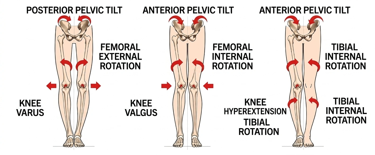
When the knee develops a varus alignment, the mechanical axis may shift medially.
As a result, the medial compartment may experience increased mechanical loading during weight-bearing activities.
Progressive medial compartment degeneration can therefore be associated with:
Increasing varus alignment
Medial joint-space narrowing
Osteophyte formation
Reduced knee motion
Progressive pain during walking
Importantly, the relationship can work in both directions.
Medial compartment degeneration can contribute to varus alignment, while varus alignment can further increase medial compartment loading.
This creates a potential biomechanical feedback loop.
The opposite pattern can occur in lateral compartment disease.
Progressive lateral compartment degeneration may be associated with valgus alignment, shifting mechanical loading toward the lateral side of the knee.
Therefore, clinicians should evaluate the entire mechanical axis rather than considering cartilage degeneration in isolation.
During standing and walking, the ground produces an upward force against the foot. This is known as the ground reaction force (GRF).
The position of this force relative to the knee joint center is clinically important.
When the GRF passes relatively close to the knee center, the resulting external moment may be relatively balanced.
When the force vector moves farther away from the joint center, the external moment acting on the knee increases.
This can alter compartmental loading and place greater mechanical demand on particular structures.
Therefore, evaluating lower-limb alignment and gait can provide information that cannot be obtained from a static knee examination alone.
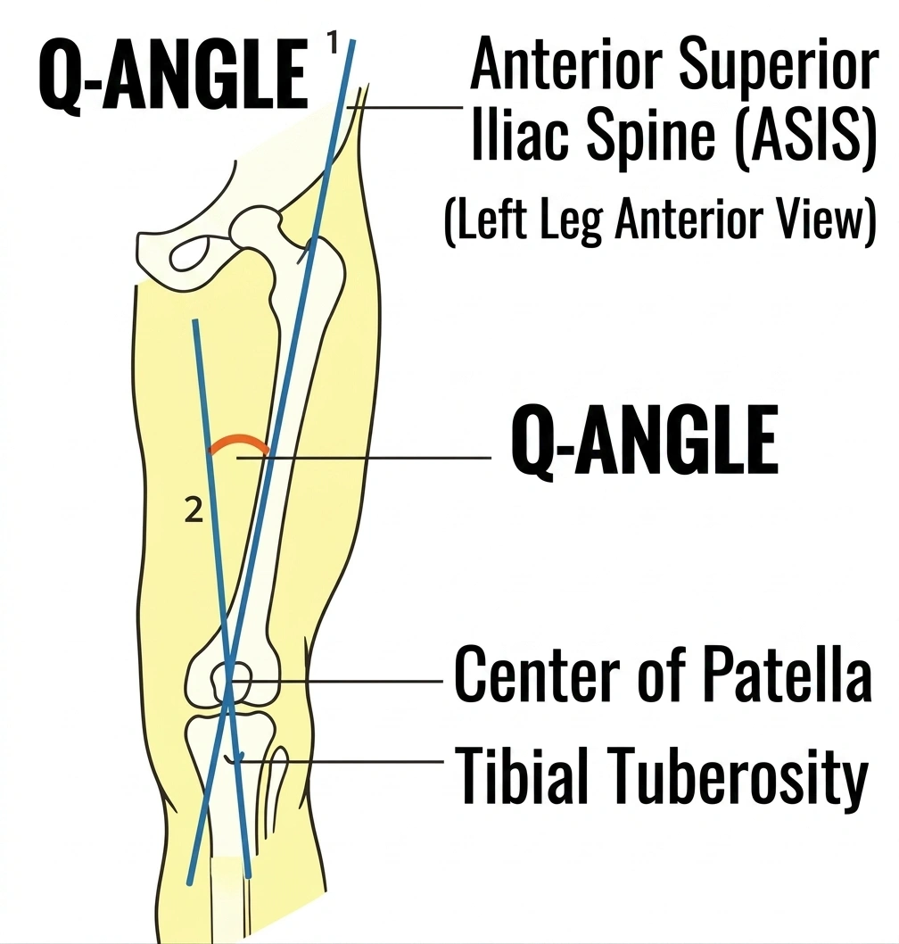
The knee does not function independently of the hip and pelvis.
The alignment between the pelvis, femur, patella and tibia can influence patellofemoral mechanics.
The Q-angle, traditionally used as an indicator of the relationship between the quadriceps mechanism and patellar tendon, is one of several parameters that may be considered during a clinical assessment.
However, the Q-angle should not be interpreted as an isolated diagnostic measurement.
Patellar tracking is also influenced by:
Quadriceps strength
Muscle balance
Patellar retinacular tension
Femoral rotation
Tibial rotation
Hip control
Foot and ankle mechanics
A comprehensive assessment therefore provides more information than relying on a single alignment measurement.
Experienced clinicians often assess whether the patella can move normally in multiple directions.
Patellar mobility may be assessed:
Superiorly
Inferiorly
Medially
Laterally
Restricted movement may suggest excessive soft-tissue tension or altered patellofemoral mechanics.
The quadriceps tendon, patellar tendon, retinaculum and surrounding soft tissues can all influence patellar mobility.
The patellofemoral joint plays an important role in knee extension and load transfer.
If the patella does not track effectively, patients may develop altered movement patterns and increased discomfort during activities such as:
Squatting
Stair climbing
Rising from a chair
Running
Repeated knee flexion and extension
Therefore, patellar assessment should be incorporated into a broader knee examination rather than being considered separately from lower-limb biomechanics.
The knee is located between the hip and foot and therefore functions as part of a kinetic chain.
Changes in foot position can influence tibial rotation and subsequently affect knee mechanics.
For example, abnormal foot posture may alter:
Tibial rotation
Ankle motion
Subtalar joint mechanics
Ground contact
Lower-limb alignment
These changes can potentially influence how forces are transmitted upward toward the knee.
This is why some patients with knee symptoms may benefit from an assessment of the foot and ankle rather than focusing exclusively on the knee.
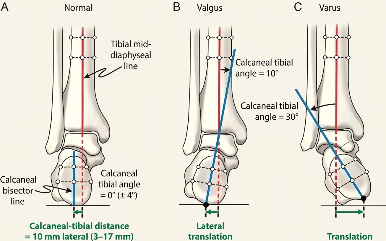
The heel and ankle complex plays an important role in lower-limb biomechanics.
The calcaneus and talus interact during weight-bearing and foot motion. Changes in calcaneal position may influence subtalar mechanics and the way the foot adapts to the ground.
Clinicians may therefore examine:
Calcaneal alignment
Subtalar motion
Rearfoot position
Ankle mobility
Foot loading pattern
Toe-off mechanics
These observations can help identify mechanical factors that may contribute to an abnormal gait.
Walking is a three-dimensional movement.
Although frontal-plane alignment is important, the knee also moves through the:
Sagittal plane
Frontal plane
Transverse plane
Therefore, correcting one plane does not automatically normalize the entire movement pattern.
For example, a patient may demonstrate relatively acceptable frontal-plane alignment but still have excessive rotational movement during walking.
A comprehensive gait assessment should therefore consider:
Evaluate:
Knee flexion and extension
Ankle dorsiflexion
Hip extension
Step length
Evaluate:
Varus or valgus alignment
Pelvic drop
Knee position during stance
Foot position
Evaluate:
Femoral rotation
Tibial rotation
Foot progression angle
Hip rotational control
The lower limb should be assessed from both directions.
In addition to looking upward from the foot toward the knee, clinicians should also evaluate the structures above the knee.
The hip and pelvis have a major influence on lower-limb alignment.
Weakness or poor motor control of the:
Gluteus medius
Gluteus maximus
Deep hip rotators
Core musculature
may contribute to abnormal femoral movement during weight-bearing.
For example, excessive femoral adduction and internal rotation during single-leg activities may alter knee mechanics.
This is particularly relevant when evaluating patients with dynamic valgus patterns.
An important clinical concept is that the knee is the middle joint between the hip and foot.
When the hip or foot cannot adequately control movement, the knee may compensate.
This can be illustrated as:
Foot → Ankle → Knee → Hip → Pelvis
or in the opposite direction:
Pelvis → Hip → Knee → Ankle → Foot
The knee may therefore become the site where excessive movement is expressed even though the original contributing factor may exist elsewhere.
This is why simply treating knee pain without identifying contributing biomechanical factors may not provide optimal long-term results for every patient.
Foot orthoses and wedge insoles can modify lower-limb loading patterns.
In selected patients with knee osteoarthritis, an appropriately designed orthosis may influence the mechanical environment of the knee and potentially reduce symptoms.
However, this approach should not be considered universally appropriate.
Depending on the patient's alignment and symptoms, an orthotic intervention may:
Modify plantar pressure
Alter tibial mechanics
Change the knee adduction moment
Improve comfort during walking
Assist movement retraining
Changing the loading pattern at the knee can simultaneously increase stress elsewhere.
For example, excessive or inappropriate wedging may increase abnormal loading around the foot or ankle.
Therefore:
An orthotic device should be prescribed according to the patient's overall biomechanics rather than simply the direction of knee deformity.
The human body does not move in a single plane.
A patient with knee degeneration may have abnormalities involving several planes simultaneously.
For example:
Frontal plane: varus or valgus alignment
Sagittal plane: limited knee extension or ankle dorsiflexion
Transverse plane: excessive femoral or tibial rotation
If treatment corrects only one component, the patient may continue to experience abnormal movement elsewhere.
Therefore, rehabilitation should ideally integrate:
Mobility training
Strength training
Neuromuscular control
Balance training
Gait retraining
Functional movement exercises
A systematic examination can help clinicians identify the major contributors to knee degeneration.
Evaluate:
Pain location
Swelling
Range of motion
Joint-line tenderness
Crepitus
Stability
Patellar mobility
Muscle strength
Evaluate:
Varus/valgus alignment
Mechanical axis
Knee position during standing
Dynamic alignment during movement
Evaluate:
Calcaneal position
Subtalar motion
Ankle dorsiflexion
Foot posture
Plantar pressure distribution
Evaluate:
Hip range of motion
Gluteal strength
Pelvic stability
Femoral rotation
Single-leg control
Observe:
Walking
Squatting
Stair climbing
Sit-to-stand
Single-leg stance
This approach provides a more complete picture of the patient's functional biomechanics.
Clinical examination should be combined with appropriate imaging when indicated.
Weight-bearing radiographs can help evaluate:
Joint-space narrowing
Osteophytes
Varus/valgus alignment
Subchondral sclerosis
Overall mechanical axis
MRI may provide additional information about:
Articular cartilage
Menisci
Ligaments
Bone marrow lesions
Synovial abnormalities
Soft-tissue structures
However, imaging findings should always be interpreted alongside clinical symptoms and functional performance.
Structural degeneration on imaging does not necessarily correspond directly to pain severity.
Management should be individualized according to disease severity, symptoms, alignment, functional limitations and patient goals.
Potential components include:
Activity modification
Quadriceps strengthening
Hip abductor strengthening
Neuromuscular training
Balance exercises
Gait retraining
Weight management when appropriate
Foot orthoses in selected patients
Appropriate analgesic strategies
Patients with persistent symptoms or advanced structural disease may require further evaluation for:
Intra-articular treatment
Osteotomy in selected alignment-related cases
Partial knee arthroplasty
Total knee arthroplasty
The appropriate treatment depends on the individual patient's pathology rather than alignment alone.
Knee degeneration should not be viewed simply as a problem of cartilage wear.
The mechanical environment of the knee is influenced by the interaction between:
Hip + Pelvis + Knee + Ankle + Foot + Gait
Varus alignment may increase medial compartment loading, while valgus alignment may increase lateral compartment loading. At the same time, foot mechanics, hip control, patellar mobility and movement patterns may influence how these forces are transmitted.
Therefore, experienced clinical assessment should move beyond the knee itself.
A useful principle is:
Do not only ask where the patient hurts. Ask why the knee is being loaded that way.
Yes. Previous ligament, meniscal, cartilage or bone injuries can alter joint mechanics and increase the risk of post-traumatic osteoarthritis.
No. Varus alignment can be associated with greater medial compartment loading, but alignment and degeneration have a bidirectional relationship. Clinical and imaging assessment is required.
Foot posture may influence tibial rotation and lower-limb mechanics, but flat feet alone do not establish that knee osteoarthritis will develop.
Insoles cannot reverse established cartilage degeneration. In selected patients, however, they may modify loading and reduce symptoms.
Often yes. Hip and pelvic control can influence lower-limb alignment during functional activities, so strengthening and neuromuscular training may be useful components of rehabilitation.
No. Weight-bearing radiographs are often sufficient for evaluating typical osteoarthritic changes. MRI is generally reserved for situations where additional soft-tissue or structural information is clinically indicated.
Knee degeneration is a complex interaction between structural damage, alignment, joint loading, muscle function and whole-limb biomechanics.
Medial and lateral compartment loading should be evaluated together with frontal-plane alignment, while sagittal and transverse-plane movement must also be considered.
The foot, ankle, hip and pelvis can all influence knee mechanics. For this reason, a comprehensive clinical assessment should not stop at the knee joint.
For patients with persistent symptoms, progressive deformity or functional limitation, appropriate imaging and specialist evaluation are essential to determine the underlying pathology and the most suitable treatment strategy.


