
Views: 0 Author: Site Editor Publish Time: 2026-08-14 Origin: Site
Bone Cement Implantation Syndrome (BCIS) is an uncommon but potentially life-threatening complication associated with orthopedic procedures involving intramedullary instrumentation, bone cement implantation, and prosthesis insertion.
BCIS is particularly important during cemented hip arthroplasty, where sudden changes in intramedullary pressure may result in the systemic release of marrow contents and other embolic material. The resulting pulmonary vascular obstruction, acute pulmonary hypertension, right ventricular dysfunction, hypoxemia, and hypotension can progress rapidly to cardiovascular collapse.
Importantly, BCIS should not be regarded as an exclusive complication of bone cement. Similar physiological events may occur during uncemented procedures, although the incidence and severity are generally lower.
For orthopedic surgeons and anesthesiologists, the most important principle is therefore not simply recognizing a "bone cement reaction," but identifying high-risk patients, anticipating critical surgical steps, recognizing early hemodynamic deterioration, and initiating coordinated resuscitation without delay.
The term "Bone Cement Implantation Syndrome" can be misleading because clinically significant embolic and cardiovascular events may also occur during uncemented arthroplasty.
Bone cement appears to increase both the frequency and severity of these events, but intramedullary manipulation itself can generate substantial embolic loads.
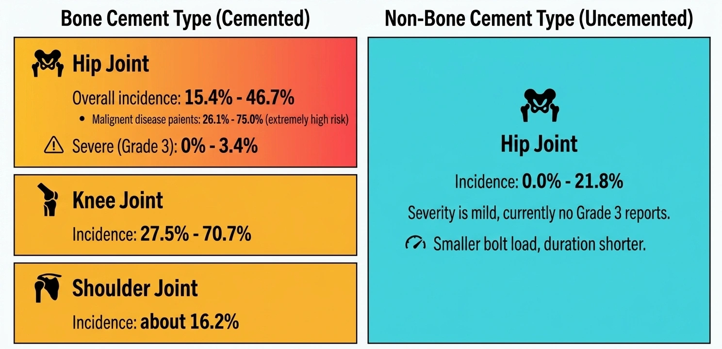
Reported incidence varies considerably depending on the definition of BCIS, patient population, surgical procedure, and severity threshold.
For cemented hip arthroplasty, reported BCIS rates have generally ranged from approximately 15% to 47%, while severe grade 3 events are much less common.
Patients undergoing arthroplasty for malignant disease may represent a particularly vulnerable population, with substantially higher reported rates of BCIS.
BCIS has also been reported during:
Cemented knee arthroplasty
Cemented shoulder arthroplasty
Revision arthroplasty
Procedures involving long femoral stems
Arthroplasty for pathological fractures
Uncemented hip arthroplasty can also produce embolic phenomena.
However, available evidence generally suggests that the embolic burden is smaller and the duration of the physiological response is shorter than with cemented implantation.
Severe grade 3 BCIS has been reported far less frequently in uncemented procedures.
The key message is:
Bone cement increases the risk of BCIS, but intramedullary manipulation and prosthesis insertion can also generate clinically important embolic events.
Therefore, appropriate monitoring should be maintained throughout the critical stages of arthroplasty rather than only during cement implantation.
The pathophysiology of BCIS is multifactorial.
The traditional explanation focused primarily on the release of bone cement components into the circulation. Current understanding, however, emphasizes intramedullary pressure, embolic load, pulmonary vascular responses, and acute right ventricular dysfunction.
Several surgical maneuvers can rapidly increase pressure inside the femoral canal, including:
Femoral canal preparation
Reaming
Cement pressurization
Cement insertion
Prosthesis insertion
Joint reduction
When intramedullary pressure exceeds venous pressure, marrow contents can be forced into the venous circulation.
The embolic material associated with BCIS may include:
Fat droplets
Bone marrow particles
Bone fragments
Air
Platelet aggregates
Cement-related particles
These materials can enter the venous system and subsequently reach the pulmonary circulation.
The resulting embolic burden may vary substantially between patients.
When embolic material reaches the pulmonary circulation, pulmonary vascular resistance may increase abruptly.
This can produce a cascade of:
Pulmonary vascular obstruction → pulmonary hypertension → right ventricular strain → reduced left ventricular filling → systemic hypotension
As pulmonary blood flow becomes impaired, ventilation-perfusion mismatch may also worsen, resulting in hypoxemia and a rapid deterioration in cardiovascular stability.
Acute right ventricular dysfunction is one of the most important mechanisms underlying severe BCIS.
The right ventricle is particularly vulnerable to a sudden increase in pulmonary vascular resistance.
As right ventricular pressure rises:
Right ventricular dilation may develop
Right ventricular contractility may deteriorate
Left ventricular preload may decrease
Cardiac output may fall
Systemic blood pressure may decline
In severe cases, this sequence can progress rapidly to cardiovascular collapse and cardiac arrest.
A patent foramen ovale (PFO) may increase the potential consequences of an acute embolic event.
Under normal conditions, increased right-sided cardiac pressure does not allow venous embolic material to enter the systemic arterial circulation.
However, when acute pulmonary hypertension causes right atrial pressure to rise sufficiently, embolic material may pass through a PFO.
This mechanism can result in paradoxical embolism, potentially affecting:
Brain circulation
Coronary circulation
Other systemic arterial territories
Consequently, patients with significant cardiopulmonary disease or known intracardiac shunts may require particularly careful perioperative risk assessment.
BCIS has traditionally been classified according to the severity of oxygen desaturation and cardiovascular instability.
Typical features may include:
Oxygen saturation below the expected range
A moderate fall in systolic blood pressure
Transient cardiovascular instability
More severe manifestations include:
Marked oxygen desaturation
Significant hypotension
Sudden loss of consciousness
Severe cardiovascular instability
Grade 3 represents the most severe form and is characterized by:
Cardiovascular collapse requiring cardiopulmonary resuscitation.
This classification is useful because it helps distinguish transient physiological changes from clinically significant cardiovascular deterioration.
Absolute blood-pressure thresholds may not adequately reflect the severity of deterioration in elderly or medically complex patients.
For example, a patient with a low baseline blood pressure may experience a clinically important reduction in perfusion despite remaining above a conventional absolute threshold.
Relative changes in mean arterial pressure (MAP) can therefore provide additional information.
A sudden substantial decline from the patient's baseline should be treated as an important warning sign, particularly when it occurs during femoral canal preparation, cementation, prosthesis insertion, or joint reduction.
Risk assessment should consider both patient-related factors and procedure-related factors.
Important risk factors include:
Advanced age
Poor overall physiological reserve
High ASA classification
Cardiovascular disease
Heart failure
Pulmonary hypertension
Significant respiratory disease
Malignancy
Pathological fracture
Poor bone quality
Pre-existing cardiopulmonary dysfunction
Patients undergoing arthroplasty for metastatic or other malignant bone disease may be particularly vulnerable because of both the underlying disease and the extensive intramedullary involvement.
Patients with limited cardiovascular reserve may tolerate an acute increase in pulmonary vascular resistance poorly.
Particular attention should be paid to patients with:
Congestive heart failure
Significant coronary artery disease
Pulmonary hypertension
Right ventricular dysfunction
Significant arrhythmias
Known intracardiac shunts
Surgical factors associated with increased risk may include:
Cemented femoral stem implantation
Long-stem prostheses
Pathological fractures
Extensive intramedullary preparation
High-pressure cement insertion
Extensive canal instrumentation
Revision procedures in selected patients
Risk assessment should therefore be performed before surgery rather than only after hemodynamic deterioration occurs.
BCIS does not occur randomly throughout the operation.
Clinicians should pay particular attention to several critical surgical steps.
Reaming and instrumentation can substantially increase intramedullary pressure and mobilize marrow contents.
This is an important period for close communication between the surgical and anesthesia teams.
Cement introduction and pressurization can produce a rapid increase in intramedullary pressure.
This is one of the most recognizable warning points for BCIS.
Insertion of the femoral stem may further displace intramedullary contents and generate embolic material.
Although often overlooked, reduction of the hip can also coincide with hemodynamic deterioration.
Therefore, monitoring should remain vigilant until the major intramedullary and reduction-related surgical maneuvers have been completed.
Early recognition can be lifesaving.
One of the most useful intraoperative indicators is a sudden reduction in end-tidal carbon dioxide (ETCO₂), particularly when it occurs together with hypotension or oxygen desaturation.
Other warning signs may include:
Sudden hypotension
Reduced SpO₂
Unexplained tachycardia or bradycardia
New cardiac arrhythmias
Increased airway pressure
Sudden reduction in cardiac output
Cardiovascular instability
Loss of consciousness in patients receiving regional anesthesia
A rapid decline in ETCO₂ may reflect a sudden reduction in pulmonary blood flow and should prompt immediate assessment for serious embolic or cardiopulmonary complications.
Transesophageal echocardiography (TEE/TOE) can provide real-time information about the right heart, pulmonary circulation, and intracardiac embolic load.
This may be particularly valuable in patients considered to be at very high risk.
TEE can help identify:
Right atrial embolic material
Right ventricular dilation
Right ventricular dysfunction
Acute pulmonary hypertension
Abnormal interventricular septal motion
Potential paradoxical embolization
The visualization of embolic material within the right heart can provide important confirmation that a major embolic event has occurred.
However, TEE should complement rather than replace continuous standard hemodynamic and respiratory monitoring.
Prevention begins before cement is introduced.
The overall objective is to reduce:
Intramedullary pressure + marrow extrusion + embolic burden + cardiopulmonary stress
Several surgical strategies may help achieve this objective.
The femoral canal can be carefully cleaned using appropriate suction and irrigation techniques.
The goal is to remove:
Marrow debris
Fat
Bone fragments
Blood
Other intramedullary material
Reducing the amount of loose material within the canal may help reduce the embolic burden generated during subsequent cementation and stem insertion.
Vacuum mixing can reduce the incorporation of air into bone cement and improve cement handling characteristics.
It may therefore contribute to reducing the amount of gaseous and particulate material generated during cement implantation.
Cement should be introduced using an appropriate technique and with careful attention to pressurization.
In high-risk patients, aggressive or uncontrolled cement pressurization should be avoided.
An appropriately selected distal cement restrictor can help establish controlled cementation and optimize cement pressurization.
Its use should follow the surgeon's established cementing technique and the characteristics of the femoral canal.
In selected cases, femoral venting may reduce intramedullary pressure.
However, creating a vent also introduces potential risks, including structural weakening of the femur.
Therefore, venting should be considered selectively rather than used automatically in every patient.
One of the most practical methods for reducing the impact of BCIS is simple but often underestimated:
communication.
Before a high-risk step, the surgeon should clearly notify the anesthesia team.
For example:
"Cement preparation."
"Cement insertion."
"Stem insertion."
"Preparing for reduction."
This allows the anesthesia team to increase vigilance immediately before the highest-risk surgical moments.
For high-risk patients, a formal BCIS team time-out can be incorporated into the surgical workflow.
The team should confirm:
Patient-specific risk factors
Planned implant type
Cemented versus uncemented strategy
Hemodynamic monitoring
Vascular access
Availability of vasoactive medications
Emergency resuscitation equipment
Escalation plan if cardiovascular collapse occurs
When sudden cardiovascular or respiratory deterioration occurs during a critical stage of arthroplasty, BCIS should be considered immediately.
The combination of:
Sudden hypotension
Rapid reduction in ETCO₂
Oxygen desaturation
New cardiac arrhythmia
Reduced cardiac output
Right ventricular dysfunction
Cardiovascular collapse
should trigger an immediate BCIS emergency response.
The timing of deterioration is particularly important. Events occurring immediately after femoral canal instrumentation, cement insertion, prosthesis implantation, or joint reduction should raise a high index of suspicion.
The team should rapidly ask:
“What happened immediately before the hemodynamic collapse?”
If the deterioration coincides with intramedullary manipulation or cementation, BCIS should be considered alongside other causes of acute intraoperative cardiovascular collapse.
The first priority is to maintain adequate oxygenation and ventilation.
In patients with suspected BCIS and hypoxemia, high-concentration supplemental oxygen should be administered immediately.
Continuous monitoring should include:
SpO₂
ETCO₂
Airway pressure
Respiratory rate
Arterial blood gases when clinically indicated
A sudden reduction in ETCO₂ is particularly important because it may indicate a major reduction in pulmonary blood flow.
If oxygenation cannot be maintained with conventional oxygen therapy, assisted or controlled mechanical ventilation may be required.
Ventilatory management should take into account the possibility of acute right ventricular dysfunction. Excessive intrathoracic pressure can further compromise venous return and right ventricular preload.
Therefore, ventilation should be carefully titrated according to the patient's hemodynamic status.
Severe BCIS can rapidly produce profound hypotension.
The initial objective is to restore adequate systemic perfusion while avoiding interventions that may further increase right ventricular stress.
For patients at high risk of severe BCIS, appropriate intravenous access should be established before the critical surgical stage.
In patients with major hemodynamic instability, invasive arterial pressure monitoring can provide continuous information about:
Blood pressure
MAP
Response to vasoactive therapy
Hemodynamic trends
Central venous access may be appropriate in selected critically ill patients when advanced vasoactive support is anticipated.
When hypotension is severe, vasoactive medications may be required.
Norepinephrine is commonly considered a first-line vasopressor for profound vasodilatory or mixed shock with hypotension, because it can restore systemic vascular pressure while supporting coronary perfusion.
Other vasoactive agents may be considered depending on:
Heart rate
Cardiac function
Severity of hypotension
Right ventricular performance
Institutional protocols
The choice of vasopressor should therefore be individualized rather than based on a single universal drug algorithm.
Volume resuscitation may be necessary when preload is inadequate, but indiscriminate fluid administration should be avoided.
This is particularly important when acute right ventricular failure is present.
Excessive volume can increase right ventricular dilation and worsen ventricular interdependence, potentially reducing left ventricular filling and systemic cardiac output.
The practical goal is:
Restore effective circulation without overloading the right ventricle.
Acute right ventricular failure is a central feature of severe BCIS.
The sudden increase in pulmonary vascular resistance creates a major pressure burden for the right ventricle.
The process can be summarized as:
Pulmonary vascular obstruction → increased RV afterload → RV dilation → reduced LV filling → decreased cardiac output → systemic hypotension
As this cycle progresses, coronary perfusion of the right ventricle may deteriorate further.
This creates a potentially self-amplifying process of:
Hypotension → reduced coronary perfusion → worsening RV function → further hypotension
Therefore, maintaining adequate systemic arterial pressure is an important component of resuscitation.
Bedside echocardiography may reveal:
Right ventricular dilation
Reduced RV systolic function
Interventricular septal flattening
Elevated pulmonary pressures
Reduced left ventricular filling
Intracardiac embolic material in selected cases
These findings can help distinguish acute right ventricular failure from other causes of intraoperative hypotension.
In a patient who is hemodynamically unstable during surgery, diagnostic evaluation should occur without unnecessarily delaying resuscitation.
The diagnosis of BCIS is primarily clinical and is supported by the timing of the event, physiological changes, and imaging findings.
Important parameters include:
ETCO₂
SpO₂
Blood pressure
ECG
Heart rate
Airway pressure
Urine output
Arterial blood gas results when indicated
A sudden fall in ETCO₂ accompanied by hypotension is particularly concerning.
ECG may show signs of acute right heart strain.
Classical findings such as S1Q3T3 can occur in acute pulmonary embolic events, although they are neither sufficiently sensitive nor specific to confirm BCIS.
Therefore, a normal ECG does not exclude BCIS, and an abnormal ECG should always be interpreted in the context of the entire clinical picture.
Point-of-care echocardiography can provide rapid information during cardiovascular collapse.
It may identify:
Acute right ventricular enlargement
Reduced RV function
Pulmonary hypertension
Abnormal ventricular interaction
Reduced left ventricular filling
Intracardiac embolic material
Transesophageal echocardiography can be particularly useful during surgery because it can provide continuous imaging without requiring the patient to leave the operating room.
However, the absence of visible embolic material does not exclude BCIS.
BCIS should not be diagnosed solely because hypotension occurs after cement implantation.
The team should also consider other potentially life-threatening causes, including:
Major hemorrhage
Myocardial infarction
Severe arrhythmia
Anaphylaxis
Tension pneumothorax
Massive pulmonary thromboembolism
High spinal or neuraxial block
Severe hypovolemia
Acute myocardial dysfunction
The timing of the event and the presence of intramedullary embolic phenomena can help establish the most likely diagnosis.
Management becomes particularly complex when severe BCIS resembles massive pulmonary embolism.
Anticoagulation or thrombolytic therapy should not be administered automatically solely on the basis of intraoperative hypotension.
The potential benefits must be weighed against the substantial bleeding risk associated with major orthopedic surgery.
Anticoagulation may be appropriate when clinically significant thromboembolic disease is strongly suspected or confirmed and when the patient's bleeding risk permits.
However, recent surgery, active bleeding, and extensive tissue trauma may significantly alter the risk-benefit calculation.
Systemic thrombolysis may be considered in selected patients with life-threatening massive pulmonary embolism and persistent hemodynamic collapse when the expected benefit outweighs the bleeding risk.
In the immediate postoperative orthopedic setting, this decision requires multidisciplinary discussion whenever the patient's condition allows.
The management should involve appropriate specialists, such as:
Anesthesiology
Orthopedic surgery
Critical care
Cardiology
Interventional radiology
Cardiothoracic surgery
When severe BCIS progresses to refractory hypoxemia, cardiogenic shock, or cardiac arrest despite conventional resuscitation, advanced mechanical support may be considered.
Venoarterial extracorporeal membrane oxygenation (VA-ECMO) can provide temporary circulatory and respiratory support in carefully selected patients with refractory cardiopulmonary failure.
Potential indications may include:
Refractory cardiogenic shock
Persistent severe hypoxemia
Cardiac arrest refractory to conventional resuscitation
Severe acute right ventricular failure
ECMO should be regarded as an advanced rescue strategy rather than routine BCIS treatment.
Early recognition is essential because ECMO requires appropriate personnel, equipment, and institutional expertise.
If imaging demonstrates a large central thromboembolic obstruction and the patient's condition remains unstable, advanced intervention may be considered.
Depending on the clinical scenario and available expertise, options can include:
Catheter-directed thrombectomy
Mechanical thrombectomy
Surgical embolectomy
Other institution-specific advanced rescue procedures
The choice depends on the underlying mechanism.
A key distinction is that BCIS may involve mixed marrow, fat, platelet, air, and cement-related embolic material, rather than a conventional thrombus alone.
Therefore, treatment should be based on the complete clinical and imaging assessment.
Patients who experience significant BCIS should generally receive continued critical-care observation even after initial stabilization.
Postoperative monitoring may include:
Continuous blood pressure monitoring
ECG
Oxygen saturation
Respiratory status
Urine output
Serial blood gas analysis
Echocardiographic reassessment when indicated
The team should remain alert for:
Persistent pulmonary hypertension
Right ventricular dysfunction
Recurrent hypotension
Hypoxemia
Arrhythmias
Acute kidney injury
Neurological complications
Secondary organ dysfunction
The severity of the initial BCIS event should guide the intensity and duration of postoperative monitoring.
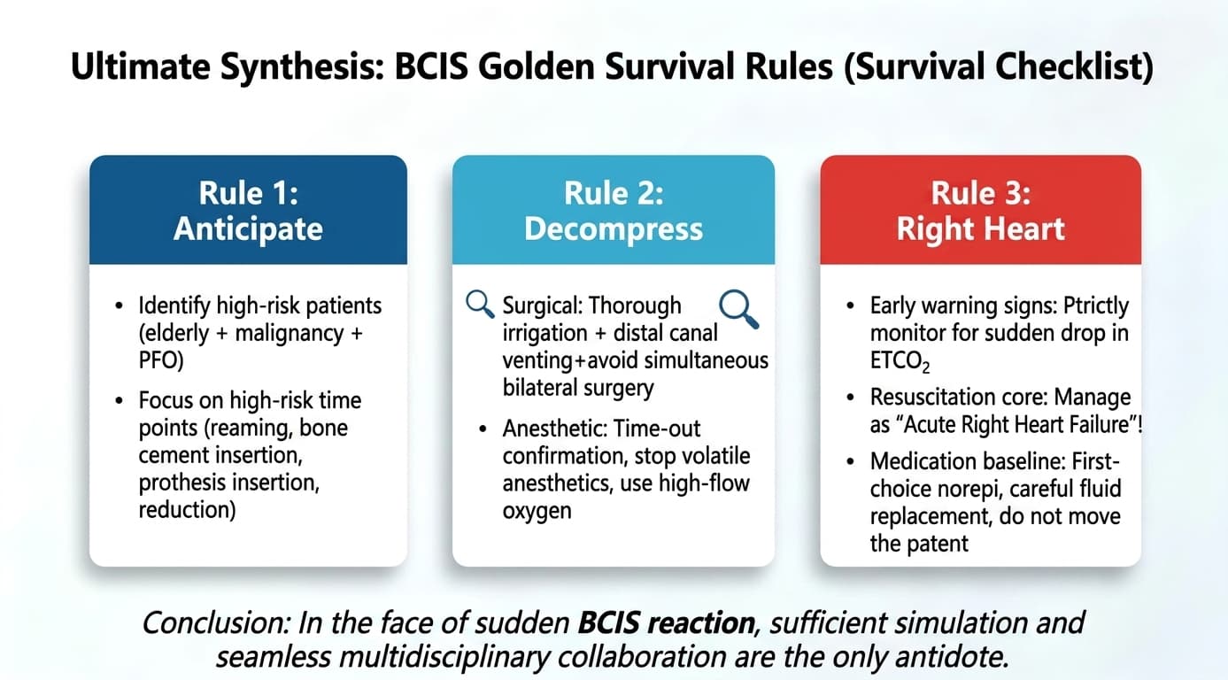
A practical BCIS response can be summarized into three principles:
Identify high-risk patients before surgery.
Pay particular attention to:
Advanced age + cardiopulmonary disease + malignancy + poor physiological reserve
Also identify high-risk procedures such as:
Cemented femoral arthroplasty
Long-stem implantation
Pathological fracture surgery
Extensive femoral canal preparation
During surgery:
Minimize unnecessary intramedullary pressure
Thoroughly clean the femoral canal
Use appropriate cementing techniques
Consider distal cement restrictors when indicated
Consider selective venting when appropriate
Avoid uncontrolled cement pressurization
Maintain close surgeon-anesthesiologist communication
When sudden deterioration occurs:
Recognize the possibility of acute right ventricular failure.
Watch for:
Sudden ETCO₂ reduction
Hypotension
Hypoxemia
Right ventricular dilation
Reduced cardiac output
Then:
Support oxygenation → restore systemic perfusion → evaluate RV function → identify the cause → escalate treatment when necessary.
BCIS should not be viewed simply as an adverse reaction to bone cement.
It is better understood as a complex perioperative cardiopulmonary syndrome associated with intramedullary manipulation and embolization.
This change in perspective has practical implications.
First, clinicians should remain vigilant even during uncemented arthroplasty.
Second, prevention should begin before cement implantation rather than after hemodynamic instability appears.
Third, the anesthesia and surgical teams should share responsibility for identifying critical moments.
Finally, the management of severe BCIS should focus on maintaining oxygenation and systemic perfusion while recognizing the central role of acute right ventricular dysfunction.
Bone Cement Implantation Syndrome is an acute perioperative cardiopulmonary syndrome characterized by hypoxemia, hypotension, cardiovascular instability, or cardiac arrest occurring around major intramedullary orthopedic procedures, particularly cemented arthroplasty.
Yes.
Although cemented arthroplasty is associated with a higher incidence and greater severity, clinically significant embolic and cardiopulmonary events can also occur during uncemented procedures.
Important surgical stages include:
Femoral canal preparation
Cement insertion
Prosthesis insertion
Joint reduction
The greatest vigilance is required during and immediately after these steps.
A sudden reduction in ETCO₂, especially when accompanied by hypotension and oxygen desaturation, is an important warning sign.
However, no single parameter is diagnostic by itself.
Pulmonary embolization can abruptly increase pulmonary vascular resistance.
The right ventricle must suddenly pump against this increased resistance, potentially resulting in acute RV dilation, reduced cardiac output, systemic hypotension, and cardiovascular collapse.
The risk cannot always be eliminated, but it can be reduced through:
Careful patient selection
Preoperative risk assessment
Appropriate cementing techniques
Intramedullary canal cleaning
Pressure control
Effective team communication
Appropriate intraoperative monitoring
No.
TEE is an advanced monitoring modality and is generally most relevant to selected high-risk patients or situations requiring detailed intraoperative assessment of embolic load and right heart function.
No.
Intraoperative hypotension has many potential causes.
BCIS should be considered particularly when cardiovascular deterioration occurs around intramedullary manipulation or cementation and is accompanied by features such as hypoxemia, ETCO₂ reduction, or acute right ventricular dysfunction.
Bone Cement Implantation Syndrome remains one of the most serious cardiopulmonary complications associated with hip and other major joint arthroplasty procedures.
Its clinical presentation can evolve within seconds, making anticipation and early recognition more important than delayed diagnosis.
The key principles are straightforward:
Identify high-risk patients.
Recognize critical surgical moments.
Reduce intramedullary pressure and embolic load.
Monitor ETCO₂ and hemodynamic changes closely.
Recognize acute right ventricular failure.
Restore oxygenation and systemic perfusion rapidly.
Escalate to advanced cardiopulmonary support when conventional treatment fails.
Most importantly, BCIS management should be multidisciplinary. Orthopedic surgeons, anesthesiologists, intensivists, cardiologists, and other specialists should have a shared understanding of the risk and an agreed emergency response pathway.
For high-risk arthroplasty, preparation is not an optional extra—it is part of the treatment strategy.
Donaldson AJ, Thomson HE, Harper NJ, Kenny NW. Bone cement implantation syndrome. Br J Anaesth. 2009;102(1):12-22.
Olsen F, Kotynek M, Wretenberg P, et al. Bone cement implantation syndrome in cemented hemiarthroplasty for femoral neck fracture. Acta Orthop. 2020.
Association of Anaesthetists. Safety guideline: reducing the risk from cemented hemiarthroplasty for hip fracture. Anaesthesia.
National Institute for Health and Care Excellence (NICE). Hip fracture management and perioperative care guidance.
British Orthopaedic Association. Guidance and resources relating to hip arthroplasty and perioperative safety.
American Society of Anesthesiologists. Practice guidance and perioperative considerations for patients undergoing major orthopedic surgery.
Note: BCIS is a potentially fatal perioperative emergency. The specific resuscitation strategy, vasoactive therapy, anticoagulation, thrombolysis, ECMO, and surgical or catheter-based intervention should be determined by the treating multidisciplinary team according to the patient's clinical condition, bleeding risk, institutional protocols, and available expertise.


